The intricate dance between our nervous system and our muscles is fundamental to every movement we make, from a subtle facial expression to a powerful athletic feat. This communication is electrical in nature, and by understanding and measuring this electrical activity, we gain profound insights into muscle function, health, and disease.
The Electrical Graph of Muscle Activity: Unveiling the Electromyogram (EMG)
The electrical activity generated by muscle fibers as they contract can be visualized and analyzed through an electromyogram (EMG). Essentially, an EMG is a graphical representation of the electrical potential produced by skeletal muscles. When a motor neuron transmits a signal to a muscle fiber, it triggers a series of electrical events that culminate in muscle contraction. This process begins with the depolarization of the sarcolemma (the muscle cell membrane) at the neuromuscular junction. This depolarization spreads along the sarcolemma and into the transverse tubules (T-tubules), initiating the release of calcium ions from the sarcoplasmic reticulum. The influx of calcium ions facilitates the interaction between actin and myosin filaments, leading to muscle shortening – the contraction.
The electrical signals generated during these events are what an EMG captures. At the cellular level, individual muscle fibers exhibit a resting membrane potential, a negative charge inside the cell relative to the outside, due to the differential distribution of ions. When stimulated, ion channels open, allowing sodium ions to rush into the cell, causing the membrane potential to become positive – this is the action potential. This rapid electrical change propagates along the muscle fiber.
When an electrode is placed on the skin over a muscle, it can detect the summation of these individual muscle fiber action potentials within the vicinity of the electrode. The EMG machine amplifies these tiny electrical signals and displays them as waveforms. The characteristics of these waveforms provide valuable diagnostic information.
Key Features of the EMG Graph:
- Baseline/Resting Activity: In a relaxed muscle, there should be minimal or no electrical activity detected by the electrodes. The baseline on the EMG graph will appear relatively flat. Any significant electrical activity during relaxation could indicate abnormal muscle firing patterns, such as spontaneous discharges or fasciculations (involuntary muscle twitches).
- Insertion Activity: Upon inserting a needle electrode into the muscle (in needle EMG), a brief burst of electrical activity is often recorded as the needle irritates the muscle fibers. This is normal and subsides quickly.
- Motor Unit Potentials (MUPs): When a muscle is voluntarily contracted, motor units begin to activate. A motor unit consists of a single motor neuron and all the muscle fibers it innately innervates. The electrical activity from a single activated motor unit, detected by a nearby electrode, is termed a Motor Unit Potential. On the EMG, MUPs typically appear as distinct, brief, biphasic or triphasic waveforms.
- Amplitude: The height of the MUP waveform is related to the number of muscle fibers within the motor unit and the synchrony of their depolarization. Larger amplitudes generally indicate more muscle fibers innervated by that motor neuron or a greater degree of synchronized firing.
- Duration: The length of the MUP waveform is influenced by the conduction velocity of the action potentials along the muscle fibers.
- Shape: The morphology (shape) of the MUP can also be altered in various neuromuscular disorders.
- Interference Pattern: As the muscle contraction strength increases, more motor units are recruited to fire, and the firing rate of already recruited motor units increases. The EMG signal then becomes a complex summation of MUPs from numerous motor units. This results in a dense, irregular pattern of waveforms, often described as an “interference pattern.”
- Full Interference Pattern: In a maximally contracted muscle, the EMG signal is so dense with overlapping MUPs that individual waveforms are difficult to discern. This indicates that all available motor units are recruited and firing at their maximum rate.
- Reduced Interference Pattern: In conditions where motor units are lost (e.g., motor neuron disease), fewer motor units are available to contribute to the contraction. Even with maximal effort, an individual may not be able to achieve a “full” interference pattern, instead displaying a “reduced” or “paucity” of MUPs.
- Recruitment: The gradual increase in the EMG signal amplitude as a muscle contraction strength is progressively increased is a key indicator of motor unit recruitment. This phenomenon will be discussed in further detail.
- Abnormal EMG Findings: Deviations from these normal patterns can indicate a range of neuromuscular pathologies. For instance, spontaneous activity like fibrillations (involuntary firing of single muscle fibers) or positive sharp waves suggest denervation (damage to the nerve supplying the muscle). Myotonic discharges, characterized by a waxing and waning amplitude and frequency, are indicative of myotonia, a condition of prolonged muscle contraction.
The EMG provides a dynamic picture of muscle electrical activity, allowing clinicians to assess nerve and muscle integrity, diagnose neuromuscular disorders, and monitor recovery.
Applying Electrodes at Appropriate Body Muscles: The Art and Science of Signal Acquisition
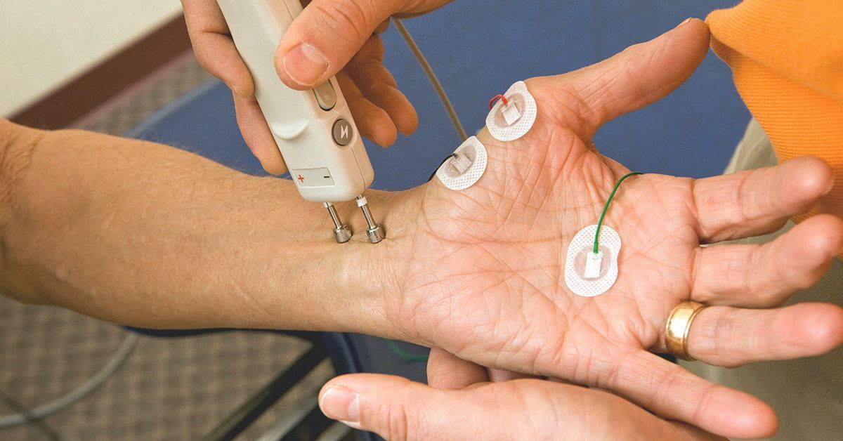
The accurate interpretation of an EMG signal hinges on the proper application of electrodes. Electrodes serve as transducers, converting the electrical signals generated by muscle activity into a readable format for the EMG machine. The choice of electrode type and their placement are critical for obtaining meaningful and diagnostically relevant data.
Types of Electrodes:
- Surface Electrodes (Suction or Adhesive): These are most commonly used for routine EMG examinations and for monitoring muscle activity during physical therapy or rehabilitation. They are placed on the skin overlying the muscle of interest.
- Advantages: Non-invasive, easy to apply, relatively comfortable for the patient.
- Disadvantages: Detect signals from a larger area, making it harder to isolate individual motor unit potentials. Signal can be affected by skin impedance, sweat, and movement artifacts. The depth of detection is limited.
- Needle Electrodes (Concentric Needle or Single-Fiber Needle): These are inserted directly into the muscle tissue.
- Concentric Needle Electrodes (CNEs): Consist of a fine needle with a central recording wire surrounded by an outer cannula, which acts as a reference electrode. They are excellent for recording motor unit potentials from a small, localized area.
- Single-Fiber Needle Electrodes (SFNEs): Have a much smaller recording surface and are designed to record from a single muscle fiber. They are invaluable for detecting subtle abnormalities in neuromuscular transmission, such as those seen in myasthenia gravis.
- Advantages: Provide a more precise assessment of individual motor units and muscle fiber activity. Can detect pathology deep within the muscle.
- Disadvantages: Invasive, can cause discomfort or pain, carry a small risk of infection or bleeding.
Principles of Electrode Placement:
The goal of electrode placement is to maximize the detection of the electrical signals from the target muscle while minimizing interference from other muscles or electrical noise.
- Identify the Muscle of Interest: Anatomical knowledge is crucial. The clinician must be able to accurately palpate and identify the muscle they intend to study. Surface landmarks, bony prominences, and the direction of muscle fibers are key guides.
- Skin Preparation: For surface electrodes, the skin should be cleaned with alcohol to remove oils and dead skin cells, ensuring good electrical contact and reducing impedance. For needle electrodes, the skin is typically cleaned with an antiseptic solution.
- Surface Electrode Placement:
- Target Muscle: The active electrode (recording electrode) is placed directly over the belly of the target muscle, typically midway between its origin and insertion, or over the area where the muscle is most prominent during contraction.
- Reference Electrode: The reference electrode is usually placed over a non-muscular bony prominence (e.g., elbow, wrist, ankle) or on an adjacent muscle that is not expected to be active during the test. This helps to minimize common-mode noise.
- Ground Electrode: A ground electrode is often used between the active and reference electrodes to further reduce electrical interference from external sources. The placement of the ground electrode can vary, but it should be somewhere in the limb being tested, away from the muscles of interest.
- Inter-electrode Distance: For surface electrodes, the distance between the active and reference electrodes can influence the signals detected. A smaller distance generally favors recording from superficial muscle fibers, while a larger distance can capture signals from deeper structures.
- Needle Electrode Placement:
- Muscle Belly Insertion: The needle electrode is inserted into the muscle belly, aiming for the center of the muscle mass where motor unit activity is likely to be robust.
- Orientation: The needle should generally be inserted parallel to the direction of the muscle fibers to optimize the detection of propagating action potentials.
- Movement and Repositioning: During needle EMG, the electrode may be moved slightly within the muscle to sample different areas and identify different motor unit potentials. The clinician listens to the auditory feedback from the EMG machine as they move the needle.
- Minimizing Artifacts:
- Movement Artifact: Patient movement, electrode cable movement, or loose electrode connections can introduce electrical noise. Patients are instructed to remain still during the recording.
- Electrical Interference: External electrical devices can interfere with EMG signals. Testing rooms are often shielded to minimize such interference.
- Sweating: Excessive sweating can create a low-resistance path for current, altering signal quality with surface electrodes.
The precise placement of electrodes, guided by anatomical knowledge and an understanding of the electrical signals being measured, is fundamental to obtaining accurate and interpretable EMG data.
The Motor Unit: The Fundamental Building Block of Muscle Control
The motor unit is the smallest functional unit of the neuromuscular system. It is the fundamental “switch” that controls muscle contraction. A motor unit consists of a single somatic motor neuron originating in the spinal cord or brainstem and all of the individual muscle fibers that this neuron innervates. When a motor neuron fires an action potential, it triggers a simultaneous contraction of all the muscle fibers it supplies.
Characteristics of a Motor Unit:
- Size Principle: Motor units are not all alike. They can be broadly categorized into “slow-twitch” (Type S) and “fast-twitch” (Type F) units, with intermediate types also existing.
- Slow-Twitch (Type S) Motor Units: These are typically small motor units, innervated by smaller, more excitable motor neurons. They have a slower conduction velocity and innervate a relatively small number of muscle fibers (e.g., tens to a few hundred). They are recruited first during voluntary contractions and fatigue resistant. They are primarily used for sustained, low-intensity activities like maintaining posture.
- Fast-Twitch (Type F) Motor Units: These are larger motor units, innervated by larger, less excitable motor neurons. They have a faster conduction velocity and innervate a much larger number of muscle fibers (e.g., hundreds to thousands). They are recruited later during contractions and generate more force but fatigue more quickly. They are crucial for rapid, powerful movements like jumping or sprinting.
- Force Output: The force produced by a single motor unit is relatively consistent. However, the total force generated by a muscle is the sum of the forces produced by all its active motor units.
- All-or-None Principle: A motor neuron, once it reaches its firing threshold, will generate an action potential, and all the muscle fibers it innervates will contract. If the stimulus is below the threshold, no action potential is generated, and no muscle fibers are activated.
- Innervation Ratio: This refers to the number of muscle fibers innervated by a single motor neuron. For fine motor control (e.g., in the fingers or eyes), the innervation ratio is low (a few muscle fibers per neuron), allowing for precise adjustments in force. For gross motor control (e.g., in the leg muscles), the innervation ratio is high (many muscle fibers per neuron), enabling the generation of powerful contractions.
The concept of the motor unit is central to understanding how we control the force and precision of our movements.
The Motor Unit Recruitment Phenomenon: Graduating Muscle Force
Motor unit recruitment is the process by which the nervous system calls upon more and more motor units to contract in order to increase the overall force generated by a muscle. This phenomenon is governed by the size principle, a fundamental concept in motor control.
The Size Principle Explained:
The size principle dictates that motor neurons are recruited in an orderly fashion, based on their size, which correlates with their excitability.
- Threshold of Recuitment (TR): Each motor neuron has a specific activation threshold – the level of excitatory input required to trigger an action potential. Smaller motor neurons have lower activation thresholds, meaning they are more easily excited and require less input to fire. Larger motor neurons have higher activation thresholds and require more significant excitatory input.
- Orderly Recruitment: When the nervous system decides to increase the force of a muscle contraction, it first activates the motor neurons with the lowest activation thresholds. These are the motor neurons that innervate slow-twitch (Type S) motor units. As the demand for force increases, progressively larger motor neurons are recruited, activating fast-twitch (Type F) motor units.
- Gradual Force Increase: This orderly recruitment allows for a smooth and graded increase in muscle force. Initially, weak contractions are generated by small motor units. As stronger contractions are needed, more and larger motor units are recruited, leading to a substantial increase in force output.
- Force Modulation: The nervous system can further modulate muscle force by altering the rate coding, which is the firing frequency of the recruited motor neurons. Even after a motor unit is recruited, its firing rate can increase, leading to greater summation of muscle twitches and thus increased force. However, the force output of a muscle is primarily determined by how many motor units are recruited, and secondarily by their firing rate.
- Fatigue Considerations: The size principle also has implications for fatigue. Slow-twitch motor units, recruited first, are fatigue-resistant and can sustain activity for long periods. Fast-twitch motor units, recruited later for higher force production, fatigue more rapidly. Therefore, as a contraction becomes prolonged and demanding, the contribution of fast-twitch units diminishes due to fatigue, leading to a decrease in overall force.
Practical Implications and EMG Observations:
The motor unit recruitment phenomenon is readily observable in EMG recordings:
- Low Force Contractions: When a muscle is contracted with very low force, only a few motor unit potentials (MUPs) will be visible on the EMG, and they will appear widely spaced. This reflects the recruitment of only the smallest, most excitable motor units.
- Increasing Force: As the force of contraction increases, more MUPs will appear on the EMG. The density of MUPs on the screen will increase, and the overall amplitude of the signal will grow. This indicates the recruitment of larger motor units.
- Maximal Contraction: During maximal voluntary contraction, the EMG will display a dense “interference pattern,” where individual MUPs are largely indistinguishable due to the simultaneous activation of numerous motor units firing at high frequencies. This signifies that virtually all available motor units within the recording range of the electrode have been recruited.
- Pathological Conditions: In diseases that destroy motor neurons (e.g., Amyotrophic Lateral Sclerosis – ALS), a reduced interference pattern is observed. Even with maximum effort, fewer motor units can be recruited, resulting in a sparser EMG signal and weaker muscle strength. Conversely, certain myopathies might show increased MUPs amplitude and duration due to collateral sprouting of surviving motor neurons to reinnervate denervated muscle fibers, leading to larger motor units.
In summary, motor unit recruitment, governed by the size principle, is the elegant mechanism by which our nervous system orchestrates muscle force. The EMG, by visualizing the electrical activity of motor units, provides a direct window into this crucial physiological process, allowing for the assessment of neuromuscular function and the diagnosis of various disorders.
References
- Enoka, R. M. (2008). Neuromuscular Adaptations to Training. Human Kinetics.
- Kim, Y. (2019). Electromyography: Basic Principles and Clinical Applications. Springer.
- Malanga, G. A., Makowiec, I., & DePrince, M. L. (2017). Electrodiagnostic Medicine: A Practical Guide. Thieme.
- Preininger, B., & Kern, H. (2008). Electromyography: A practical guide for clinicians. Springer Science & Business Media.
- Ross, A., & Fredrickson, L. (2009). Clinical Electromyography. Lippincott Williams & Wilkins.


