The basal nuclei, often referred to as the basal ganglia, represent a crucial group of subcortical nuclei deeply embedded within the cerebral hemispheres. Far from being a mere motor control center, these complex structures play a pivotal role in modulating a broad spectrum of functions, including motor control, cognitive processes, emotional regulation, and procedural learning. Their intricate anatomical organization and highly interconnected neural circuits are fundamental for selecting appropriate actions, inhibiting unwanted movements, and facilitating goal-directed behaviors. A comprehensive understanding of the basal nuclei is indispensable for grasping the neurological underpinnings of various movement disorders and psychiatric conditions.
The Location and Components of Basal Nuclei
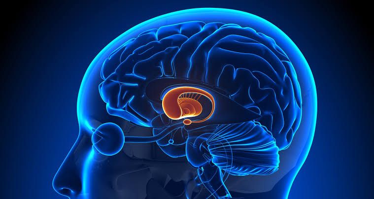
The basal nuclei are primarily located within the forebrain, specifically deep to the cerebral cortex and largely surrounding the thalamus. They are subcortical structures, meaning they lie beneath the cerebral cortex. While the term “basal ganglia” historically refers to a collection of nuclei at the base of the cerebrum, the more anatomically precise term “basal nuclei” is often preferred in modern neuroscience.
The principal components of the basal nuclei, based on their functional connectivity and role in the established circuits, include:
- Striatum: This is the primary input nucleus of the basal nuclei, receiving extensive projections from the entire cerebral cortex. It is anatomically divided into two main parts by the internal capsule:
- Caudate Nucleus: Characterized by a C-shape, it comprises a head (anteriorly, bulging into the lateral ventricle), a body, and a tail (extending posteriorly and inferiorly, ending near the amygdala). The caudate is particularly involved in cognitive and oculomotor functions.
- Putamen: A large, somewhat oval-shaped nucleus located more laterally than the caudate. The putamen is predominantly involved in motor and sensorimotor functions.
- Note: Functionally, the caudate and putamen are often considered a single unit, the “neostriatum” or simply “striatum,” due to their shared embryological origin and similar afferent connections. The term “lentiform nucleus” is an anatomical descriptor for the combined putamen and globus pallidus because of their lens-like shape.
- Globus Pallidus (Pallidum): This is the major output nucleus of the basal nuclei system, acting as a “brake” on the thalamus. It is divided into two segments:
- Globus Pallidus externa (GPe): The outer segment, involved in the indirect pathway.
- Globus Pallidus interna (GPi): The inner segment, which forms the primary output of the basal nuclei, sending inhibitory projections to the thalamus. The GPi’s function is often linked with the substantia nigra pars reticulata due to their similar output roles.
- Substantia Nigra (SN): Located in the midbrain, the substantia nigra is crucial for modulating basal nuclei activity. It is divided into two distinct parts:
- Pars Compacta (SNpc): Contains dopaminergic neurons that project predominantly to the striatum. These neurons are vital for motor control, and their degeneration is a hallmark of Parkinson’s disease.
- Pars Reticulata (SNpr): Consists of GABAergic neurons that serve as another major output pathway of the basal nuclei, projecting to the thalamus and superior colliculus. Functionally, the SNpr is considered equivalent to the GPi.
- Subthalamic Nucleus (STN): A small, biconvex nucleus situated in the diencephalon, ventral to the thalamus and medial to the internal capsule. It is the only nucleus within the basal nuclei circuit that uses an excitatory neurotransmitter (glutamate) to influence other basal nuclei components, playing a critical role in the indirect pathway.
While the amygdala and claustrum are sometimes mentioned in association with the basal nuclei due to their deep cerebral location, they are typically considered separate functional entities in the context of the classic basal nuclei circuits.
The Connections of Basal Nuclei
The functional architecture of the basal nuclei is defined by a complex network of interconnected loops, primarily involving the cerebral cortex, thalamus, and the basal nuclei components themselves. These circuits allow the basal nuclei to exert a modulatory influence over motor, cognitive, and limbic functions. The most widely accepted model describes two primary pathways that originate in the striatum and project through the globus pallidus and substantia nigra to the thalamus: the direct pathway and the indirect pathway, exquisitely balanced by dopaminergic input.
A. Corticostriatal Input: The striatum (caudate and putamen) receives a massive, topographically organized excitatory (glutamatergic) input from almost the entire cerebral cortex. Different cortical areas project to specific regions of the striatum, forming parallel loops for motor, cognitive, and limbic functions. For instance, motor and somatosensory cortices project to the putamen, while prefrontal and parietal cortices project to the caudate and anterior putamen, and limbic cortices project to the ventral striatum (including the nucleus accumbens).
B. The Direct Pathway (Go Pathway): This pathway serves to facilitate desired movements by disinhibiting the thalamus.
- Cortex to Striatum: Excitatory glutamatergic projections from the cerebral cortex activate medium spiny neurons (MSNs) in the striatum.
- Striatum to GPi/SNpr: These MSNs in the striatum are GABAergic (inhibitory) and specifically express D1 dopamine receptors. When activated, they inhibit the globus pallidus interna (GPi) and substantia nigra pars reticulata (SNpr).
- GPi/SNpr to Thalamus: The GPi/SNpr are tonically active and exert continuous inhibitory (GABAergic) control over the ventral anterior (VA) and ventral lateral (VL) nuclei of the thalamus.
- Thalamus to Cortex: Inhibition of GPi/SNpr by the striatum reduces the tonic inhibition of the thalamus. This disinhibition allows the thalamus to become more active, sending excitatory glutamatergic projections back to the motor and premotor areas of the cerebral cortex, thereby facilitating movement.
C. The Indirect Pathway (Stop Pathway): This pathway works to suppress unwanted movements by increasing inhibition of the thalamus.
- Cortex to Striatum: Excitatory glutamatergic projections from the cerebral cortex also activate a different population of MSNs in the striatum. These MSNs are GABAergic and specifically express D2 dopamine receptors.
- Striatum to GPe: Activated D2-MSNs inhibit the globus pallidus externa (GPe).
- GPe to STN: The GPe is tonically active and inhibits the subthalamic nucleus (STN). When the GPe is inhibited by the striatum, its inhibition of the STN is reduced, leading to disinhibition and activation of the STN.
- STN to GPi/SNpr: The STN sends excitatory glutamatergic projections to the GPi/SNpr.
- GPi/SNpr to Thalamus: Increased excitation of the GPi/SNpr by the STN leads to increased inhibition of the thalamus. This heightened inhibition suppresses thalamocortical activity, thereby inhibiting unwanted movements.
D. Dopaminergic Modulation by Substantia Nigra Pars Compacta (SNpc): The SNpc plays a crucial role in balancing these two pathways through its dopaminergic projections to the striatum:
- Direct Pathway: Dopamine released from SNpc acts on D1 receptors (excitatory) on direct pathway MSNs, enhancing their activity and thus promoting movement.
- Indirect Pathway: Dopamine acts on D2 receptors (inhibitory) on indirect pathway MSNs, reducing their activity and thus suppressing the indirect pathway’s inhibitory effect on movement.
In essence, dopamine from the SNpc simultaneously facilitates the direct pathway and inhibits the indirect pathway, leading to an overall enhancement of motor output and facilitation of movement.
E. Other Key Connections:
- Thalamocortical Projections (Output): The major output of the basal nuclei is relayed through the thalamus (VA/VL nuclei) back to the cerebral cortex, primarily to motor and premotor areas, but also to prefrontal and limbic cortices.
- Pallidothalamic Projections: The GPi and SNpr are the main output nuclei, sending GABAergic projections to various thalamic nuclei.
- Subthalamopallidal Projections: The STN sends glutamatergic projections to both GPe and GPi, forming a critical regulatory node.
- Brainstem Connections: The basal nuclei also have output connections to some brainstem nuclei, influencing descending motor pathways.
F. Parallel Cortical-Basal Ganglia-Thalamocortical Loops: Beyond the classic motor loop, modern neuroscience recognizes several parallel, segregated loops connecting specific cortical areas through the basal nuclei and thalamus back to the cortex. These include:
- Motor Loop: Involves sensorimotor cortex, putamen, GPi/SNpr, and motor thalamus (VA/VL), crucial for motor control.
- Associative/Cognitive Loop: Involves prefrontal cortex, caudate, GPi/SNpr, and cognitive thalamus, important for executive functions, planning, and goal-directed behavior.
- Limbic Loop: Involves anterior cingulate cortex, ventral striatum (nucleus accumbens), ventral pallidum, and limbic thalamus, mediating reward, motivation, emotion, and habit formation.
These interconnected circuits allow the basal nuclei to perform their complex functions of action selection, motor learning, and cognitive control by finely tuning the balance between facilitating desired actions and suppressing competing, unwanted ones.
Clinical Aspects Related to Basal Nuclei
Dysfunction within the intricate basal nuclei circuits is a hallmark of a wide range of neurological and psychiatric conditions, predominantly manifesting as disorders of movement, but also affecting cognition and emotion. These clinical aspects underscore the critical roles of each component and their delicate balance.
A. Movement Disorders:
- Parkinson’s Disease (PD) – Hypokinetic Disorder:
- Pathology: Characterized by the progressive degeneration of dopaminergic neurons in the substantia nigra pars compacta (SNpc). This loss of dopamine is the principal cause of motor symptoms.
- Mechanism: Reduced dopamine input to the striatum leads to an imbalance in the direct and indirect pathways.
- Direct Pathway: Less dopamine means less excitation of D1 receptors, reducing the facilitation of movement.
- Indirect Pathway: Less dopamine means less inhibition of D2 receptors, thus strengthening the indirect pathway and increasing the suppression of movement.
- The net effect is increased inhibition of the thalamus by GPi/SNpr, leading to reduced excitatory drive to the motor cortex.
- Symptoms: Bradykinesia (slowness of movement), rigidity (increased muscle tone), resting tremor (classic “pill-rolling” tremor), and postural instability. Non-motor symptoms include cognitive decline, depression, and olfactory dysfunction.
- Treatment: Levodopa (precursor to dopamine), dopamine agonists, MAO-B inhibitors, and in advanced cases, Deep Brain Stimulation (DBS) targeting the STN or GPi.
- Huntington’s Disease (HD) – Hyperkinetic Disorder:
- Pathology: An autosomal dominant neurodegenerative genetic disorder caused by an expanded CAG trinucleotide repeat in the huntingtin gene. It primarily affects the medium spiny neurons (MSNs) in the striatum, particularly those projecting to the GPe (indirect pathway).
- Mechanism: Selective degeneration of D2-receptor-expressing MSNs in the striatum effectively weakens the indirect pathway. This leads to reduced inhibition of GPe, which then leads to increased inhibition of STN. The reduced STN activity then results in less excitation of GPi/SNpr, ultimately reducing the tonic inhibition of the thalamus. The direct pathway, being relatively spared initially, becomes more dominant.
- Symptoms: Chorea (involuntary, jerky, dance-like movements), athetosis (slow, writhing movements), dystonia, cognitive decline (executive dysfunction, memory loss), and significant psychiatric symptoms (depression, irritability, psychosis).
- Treatment: Symptomatic, using dopamine-depleting agents (e.g., tetrabenazine) or antipsychotics to manage chorea. No cure currently available.
- Hemiballismus – Hyperkinetic Disorder:
- Pathology: Typically caused by a lesion (most commonly a stroke) in the subthalamic nucleus (STN), usually unilaterally.
- Mechanism: Damage to the STN removes its excitatory drive to the GPi/SNpr in the indirect pathway. This disinhibits the GPi/SNpr, leading to reduced inhibition of the thalamus on the contralateral side. The result is an overactive motor cortex and uncontrolled movements.
- Symptoms: Violent, flinging, involuntary movements of the contralateral limbs (arm and leg, or just arm).
- Treatment: Dopamine receptor blocking agents; in refractory cases, DBS or pallidotomy can be considered.
- Dystonia – Hyperkinetic Disorder:
- Pathology: A diverse group of movement disorders characterized by sustained or intermittent muscle contractions causing abnormal, often repetitive, movements, postures, or both. The underlying mechanisms are complex but often involve basal ganglia dysfunction, particularly an imbalance in the direct and indirect pathways. It can be genetic, acquired, or idiopathic.
- Mechanism: Hyperexcitability in the direct pathway or reduced inhibition from the indirect pathway, leading to abnormal co-contraction of agonist and antagonist muscles.
- Symptoms: Manifest as twisting movements, repetitive actions, or unusual postures in focal areas (e.g., cervical dystonia affecting the neck, blepharospasm affecting eyelids) or generalized throughout the body.
- Treatment: Botulinum toxin injections (for focal dystonia), oral medications (e.g., anticholinergics, benzodiazepines), and DBS (for generalized or refractory dystonia).
- Tourette Syndrome – Hyperkinetic Disorder:
- Pathology: A neurodevelopmental disorder believed to involve dysfunction in the cortico-striato-thalamo-cortical loops, with particular emphasis on dopaminergic system dysregulation.
- Mechanism: Imbalances in dopamine transmission in the striatum, leading to a reduced ability to suppress unwanted motor and vocal outputs.
- Symptoms: Characterized by multiple motor tics and at least one vocal tic, which are typically sudden, rapid, recurrent, nonrhythmic motor movements or vocalizations.
- Treatment: Behavioral therapy (e.g., Comprehensive Behavioral Intervention for Tics – CBIT), alpha-2 adrenergic agonists, and dopamine receptor blocking agents.
B. Non-Motor Clinical Aspects: Beyond movement disorders, the basal nuclei are increasingly recognized for their involvement in a range of cognitive and psychiatric conditions, reflecting the existence of parallel non-motor loops.
- Obsessive-Compulsive Disorder (OCD): Dysregulation within the associative and limbic cortico-basal ganglia-thalamocortical loops is strongly implicated. Overactivity of specific circuits linking the orbitofrontal cortex, caudate nucleus, and thalamus is thought to contribute to compulsive behaviors and intrusive thoughts.
- Addiction: The ventral striatum, particularly the nucleus accumbens, is a core component of the brain’s reward system and plays a central role in motivation, reward processing, and the formation of habits. Chronic substance abuse significantly alters the function and structure of these basal nuclei circuits, contributing to the compulsive drug-seeking and taking behaviors characteristic of addiction.
- Cognitive Impairments: Many basal nuclei disorders, such as Parkinson’s and Huntington’s disease, are associated with significant cognitive deficits, including executive dysfunction, problems with attention, working memory, and decision-making, highlighting the basal nuclei’s role in cognitive processing beyond motor control.
- Depression and Anxiety: The limbic loops involving the ventral striatum are intimately connected with mood regulation. Dysfunctions in these circuits can contribute to symptoms of depression, anxiety disorders, and apathy.
In conclusion, the basal nuclei are highly intricate subcortical structures whose precise location and diverse components underpin their critical roles in motor control, cognition, and emotion. Their complex interconnections, particularly the direct and indirect pathways balanced by dopaminergic input, serve as a sophisticated modulator of neural activity, facilitating desired actions and suppressing unwanted ones. Clinical conditions arising from basal nuclei dysfunction, ranging from debilitating movement disorders like Parkinson’s and Huntington’s diseases to significant contributions to psychiatric conditions, profoundly illustrate their indispensable role in maintaining neurological health and underscore the importance of continued research into these fascinating brain regions.
References
- Purves, D., Augustine, G. J., Fitzpatrick, D., Katz, L. C., LaMantia, A. S., McNamara, J. O., & Williams, S. M. (2001). Neuroscience (2nd ed.). Sinauer Associates.
- Bear, M. F., Connors, B. W., & Paradiso, M. A. (2016). Neuroscience: Exploring the Brain (4th ed.). Wolters Kluwer.
- Kandel, E. R., Schwartz, J. H., Jessell, T. M., Siegelbaum, S. A., & Hudspeth, A. J. (Eds.). (2013). Principles of Neural Science (5th ed.). McGraw-Hill Education.
- Lanciego, J. L., Luquin, N., & Obeso, J. A. (2012). Functional neuroanatomy of the basal ganglia. Movement Disorders, 27(12), 1483-1499.
- Parent, A., & Hazrati, L. N. (1995). Functional anatomy of the basal ganglia. I. The cortico-basal ganglia-thalamo-cortical loop. Brain Research Reviews, 20(1), 91-127.
- Calabresi, P., Picconi, B., Tozzi, A., & Di Filippo, M. (2014). Parkinson’s disease as a synaptic disorder. Current Opinion in Neurology, 27(4), 421-429.
- Albin, R. L., Young, A. B., & Penney, J. B. (1989). The functional anatomy of basal ganglia disorders. Trends in Neurosciences, 12(10), 366-375.
- Alexander, G. E., DeLong, M. R., & Strick, P. L. (1986). Parallel organization of functionally segregated circuits linking basal ganglia and cortex. Annual Review of Neuroscience, 9(1), 357-381.
- DeLisi, L. E., & Weinberger, D. R. (2019). The Neuroscience of Schizophrenia. In L. H. D. E. Martin (Ed.), Psychological Aspects of Neuroscience (pp. 53-73). Springer.
- Gurney, K. N., Prescott, T. J., & Redgrave, P. (2001). A computational model of action selection in the basal ganglia. I. A new functional anatomy. Biological Cybernetics, 84(6), 401-412.


