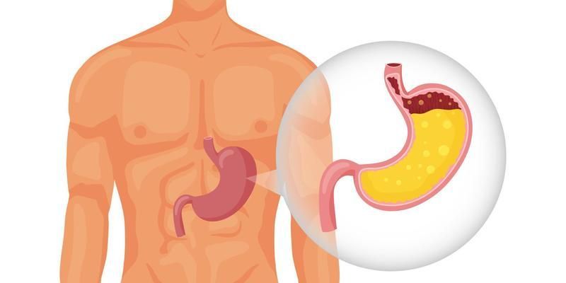
The stomach is a remarkable organ, central to the initial stages of chemical and mechanical digestion. Its ability to break down complex macromolecules, sterilize ingested food, and prepare nutrients for absorption is largely dependent on the sophisticated activity of its gastric glands. These microscopic structures secrete a potent cocktail known as gastric juice, the production of which is under precise neural and hormonal control.
Physiological Anatomy of Gastric Glands
The inner lining of the stomach, the gastric mucosa, is not a flat surface but is pocked with millions of deep indentations called gastric pits. These pits are the openings to the gastric glands, which extend down into the mucosa. While glands in different stomach regions (cardia, fundus, body, and antrum) vary slightly, the most functionally significant are the oxyntic (or gastric) glands, located in the fundus and body, which are responsible for secreting most of the gastric juice.
An oxyntic gland can be divided into three regions: the isthmus (closest to the pit), the neck, and the base. Within this structure reside several distinct and vital cell types:
- Mucous Neck Cells: Located in the neck region, these cells secrete a thin, soluble mucus that differs from the thick, viscous mucus produced by surface epithelial cells.
- Parietal (Oxyntic) Cells: Found primarily in the neck and deeper parts of the gland, these large, pyramid-shaped cells are the powerhouses of acid secretion. They produce hydrochloric acid (HCl) and Intrinsic Factor, a glycoprotein essential for vitamin B12 absorption.
- Chief (Peptic) Cells: These cells are most numerous in the base of the gland. Their primary function is to synthesize and secrete pepsinogen, the inactive precursor (zymogen) of the potent protein-digesting enzyme, pepsin.
- Enteroendocrine Cells: Scattered throughout the glands, these cells function as local hormone producers. Key examples include G-cells (mainly in the antral glands), which secrete the hormone gastrin, and D-cells, which secrete somatostatin, an inhibitory hormone.
Composition and Functions of Gastric Juice
Gastric juice is a highly acidic, watery fluid containing a mixture of substances produced by the various glandular cells. Each day, the stomach produces approximately 1.5 to 2 liters of this secretion. Its primary components and their functions are outlined below.
- Water: The primary solvent, making up over 99% of gastric juice, facilitating chemical reactions and the mixing of food into a semi-liquid paste called chyme.
- Hydrochloric Acid (HCl): Secreted by parietal cells, HCl is responsible for the extremely low pH (1.5-3.5) of gastric juice. Its functions are critical:
- Sterilization: It kills most ingested microorganisms, including bacteria and viruses, providing a crucial first line of defense against foodborne pathogens.
- Protein Denaturation: The acidic environment unfolds the complex three-dimensional structures of dietary proteins, exposing their peptide bonds to enzymatic attack.
- Enzyme Activation: HCl converts inactive pepsinogen into its active form, pepsin.
- Pepsin: As the active form of pepsinogen, pepsin is a powerful protease. It initiates protein digestion by breaking down large protein molecules into smaller polypeptides. It functions optimally in the highly acidic environment created by HCl.
- Intrinsic Factor: This glycoprotein, also secreted by parietal cells, binds to vitamin B12 in the stomach. This complex protects the vitamin from degradation and is essential for its absorption later in the terminal ileum. A deficiency of intrinsic factor leads to pernicious anemia.
- Mucus and Bicarbonate: The surface mucous cells and mucous neck cells secrete a thick layer of alkaline mucus rich in bicarbonate ions (HCO₃⁻). This forms the gastric mucosal barrier, a protective layer that neutralizes HCl at the epithelial surface, preventing the stomach from digesting itself.
- Gastric Lipase: Secreted by chief cells, this enzyme plays a minor role in initiating the digestion of triglycerides (fats), but its contribution is far less significant than that of pancreatic lipase in the small intestine.
The Mechanism of HCl Secretion
The secretion of HCl by parietal cells is an energy-intensive process that results in a hydrogen ion concentration in the gastric lumen up to three million times greater than in the blood. This feat is accomplished by the H⁺/K⁺-ATPase, commonly known as the proton pump.
- Carbon Dioxide Diffusion: Carbon dioxide from the blood diffuses into the parietal cell.
- Carbonic Acid Formation: Inside the cell, the enzyme carbonic anhydrase rapidly catalyzes the reaction of CO₂ with water (H₂O) to form carbonic acid (H₂CO₃).
- Ion Dissociation: Carbonic acid spontaneously dissociates into a hydrogen ion (H⁺) and a bicarbonate ion (HCO₃⁻).
- Proton Pumping: The H⁺/K⁺-ATPase pump, located on the apical (luminal) membrane of the cell, actively transports H⁺ into the gastric gland lumen in exchange for a potassium ion (K⁺). This is the primary active step requiring ATP.
- Chloride and Potassium Transport: The bicarbonate ion (HCO₃⁻) is transported out of the cell across the basolateral (blood-side) membrane in exchange for a chloride ion (Cl⁻) from the blood. This efflux of bicarbonate into the blood creates a temporary post-meal rise in blood pH known as the “alkaline tide.” The accumulated Cl⁻ then diffuses through channels in the apical membrane into the lumen, following the electrical gradient established by the pumping of H⁺. The K⁺ that was pumped into the cell also recycles back into the lumen via K⁺ channels.
- HCl Formation: In the lumen of the gland, the secreted H⁺ and Cl⁻ combine to form hydrochloric acid.
Regulation and Control of Gastric Juice Secretion
The secretion of gastric juice is not constant but is precisely regulated by a combination of neural and hormonal mechanisms, traditionally divided into three phases based on the location of the stimulus.
- Phase 1: Cephalic Phase: This phase is initiated by the sight, smell, thought, or taste of food, even before it enters the stomach. The central nervous system, via the vagus nerve (parasympathetic stimulation), directly stimulates parietal cells to secrete HCl and G-cells to release gastrin. This preparatory phase accounts for about 30% of the total gastric secretion in response to a meal.
- Phase 2: Gastric Phase: This is the major phase, accounting for 60% of secretion. It begins once food enters the stomach. It is stimulated by two main triggers:
- Stomach Distension: Stretching of the stomach wall activates mechanoreceptors, triggering both local (short) and vagovagal (long) neural reflexes that stimulate secretion.
- Chemical Stimuli: The presence of peptides and amino acids (from protein digestion) directly stimulates G-cells in the antrum to release the hormone gastrin into the bloodstream. Gastrin is the most potent stimulator of acid secretion, acting on parietal cells.
- Phase 3: Intestinal Phase: This phase has both an initial excitatory component and a more dominant inhibitory component. When chyme first enters the duodenum, it can briefly stimulate gastrin release. However, as the duodenum fills, particularly with acidic (pH < 2) or fatty chyme, inhibitory signals dominate to slow gastric activity. This is mediated by:
- The Enterogastric Reflex: A neural reflex that inhibits vagal stimulation.
- Hormonal Inhibition: The release of intestinal hormones such as secretin (in response to acid), cholecystokinin (CCK) (in response to fats and proteins), and somatostatin powerfully inhibits parietal cell secretion, G-cell activity, and gastric motility. This ensures the small intestine is not overwhelmed.
At the cellular level, the regulation of acid secretion is controlled by three key agonists: acetylcholine (from nerve endings), gastrin (hormone), and histamine (a paracrine substance released from nearby enterochromaffin-like cells). Somatostatin acts as the primary inhibitor. These substances bind to specific receptors on the parietal cell, modulating the activity of the proton pump and thus controlling the final output of acid.
References
- Hall, J. E. (2021). Guyton and Hall Textbook of Medical Physiology (14th ed.). Elsevier.
- Boron, W. F., & Boulpaep, E. L. (2017). Medical Physiology (3rd ed.). Elsevier.
- Johnson, L. R. (2019). Gastrointestinal Physiology (9th ed.). Mosby.
- Koeppen, B. M., & Stanton, B. A. (2018). Berne & Levy Physiology (7th ed.). Elsevier.


