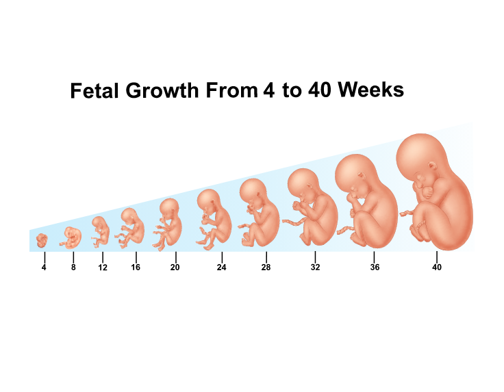
Fetal growth abnormalities (FGA) represent a spectrum of conditions where the fetus fails to achieve its genetically predetermined growth potential (Fetal Growth Restriction, FGR) or exhibits excessive growth (Macrosomia). These conditions are major determinants of perinatal morbidity and mortality worldwide, necessitating rigorous monitoring and timely intervention rooted in evidence and value-based care principles.
Explaining Fetal Growth Abnormalities and Key Definitions
Fetal growth is a dynamic process influenced by maternal health, placental function, and fetal genetic potential. Abnormalities arise when these inputs are dysregulated, leading to deviations from standard growth curves.
Defining Macrosomia and Fetal Growth Restriction (FGR)
Fetal Growth Restriction (FGR): FGR is a pathological condition where the fetus fails to achieve its growth potential, often due to placental insufficiency or adverse maternal factors. Clinically, FGR is frequently defined by an estimated fetal weight (EFW) or abdominal circumference (AC) below the 10th percentile for gestational age (Small for Gestational Age, SGA). Critically, not all SGA infants are pathologically restricted; FGR specifically implies a high risk of adverse outcomes, often diagnosed through additional evidence of fetal compromise (e.g., abnormal Doppler flow studies). FGR is further categorized as early-onset (usually pre-32 weeks, often associated with severe placental disease) or late-onset (post-32 weeks, more subtle).
Macrosomia (Large for Gestational Age, LGA): Macrosomia refers to a fetus that exhibits excessive growth. While there is no global consensus, it is typically defined as an EFW or birth weight above the 90th percentile for gestational age, or an absolute birth weight exceeding 4,000 grams (approximately 8 lbs 13 oz), irrespective of gestational age. Macrosomia is often associated with maternal conditions, most notably pre-existing or gestational diabetes mellitus.
Describing Etiologies of Abnormal Growth
The causes of FGA are multifactorial, encompassing maternal, placental, and fetal factors.
Etiologies of Fetal Growth Restriction (FGR):
- Placental Insufficiency: The most common cause, restricting the flow of oxygen and nutrients. This can result from chronic hypertension, preeclampsia, antiphospholipid syndrome, or pre-existing maternal vascular disease.
- Maternal Factors: Chronic maternal diseases (e.g., severe renal disease, cyanotic heart disease), substance abuse (smoking, alcohol, illicit drugs), chronic infections (TORCH, malaria), and extreme maternal age.
- Fetal Factors: Chromosomal abnormalities (e.g., Trisomy 13, 18), genetic syndromes, and multiple gestations.
Etiologies of Macrosomia:
- Maternal Diabetes: Poor glycemic control leads to fetal hyperglycemia and hyperinsulinemia, stimulating somatic growth (visceromegaly and increased fat deposition).
- Maternal Obesity and Excessive Gestational Weight Gain: High pre-pregnancy BMI and rapid weight gain are independently linked to LGA.
- Genetic/Constitutional Factors: Tall or larger parents often produce LGA infants.
- Post-Term Pregnancy: Extended gestation increases growth accumulation.
Effects of Socio-Economic Status (SES) and Nutrition:
Socio-economic status and nutrition significantly modulate the risk of FGA.
FGR and Low SES: Low SES is a powerful independent predictor of FGR. This connection operates through several mechanisms:
- Malnutrition: Chronic maternal caloric and micronutrient deficiencies (e.g., inadequate folate, iron) directly impair fetal growth.
- Access to Care: Delayed or inconsistent prenatal care reduces the likelihood of identifying predisposing conditions (like hypertension) or diagnosing FGR early.
- Chronic Stress: High levels of chronic stress associated with economic hardship can impact maternal physiology, increasing systemic resistance and potentially worsening placental blood flow (the “weathering” phenomenon).
Macrosomia and Nutrition/SES: While malnutrition is tied to FGR, the rise in macrosomia is often linked to the global obesity epidemic. In high-resource settings and increasingly among lower SES populations, diets high in refined sugars and saturated fats contribute to maternal obesity and uncontrolled gestational diabetes, fueling macrosomia.
Detection Methods for Fetal Growth Abnormalities
Detection requires a structured, tiered approach, prioritizing cost-effectiveness and targeted screening—the foundation of value-based care (VBC).
Standard Surveillance Methods:
- Fundal Height Measurement (SFH): A primary, low-cost screening tool. SFH measurements falling below the 10th percentile, or a lag of $\geq 3$ cm from gestational age, mandate further investigation. While relatively insensitive, its near-universal application makes it the crucial first detection step in VBC.
- Maternal Risk Assessment: Early identification of risk factors (e.g., history of FGR, chronic hypertension, diabetes) guides the need for enhanced surveillance.
Advanced Diagnostic Methods:
- Ultrasound Biometry: The gold standard for diagnosis. Serial measurements of the EFW and AC are used to plot growth trends. Early dating ultrasound is critical, as accurate gestational age is paramount for diagnosing deviations.
- Doppler Velocimetry: Essential for distinguishing pathological FGR from small non-compromised fetuses (SGA). Abnormal umbilical or middle cerebral artery Dopplers indicate placental compromise and predict adverse outcomes, justifying intervention.
- Amniotic Fluid Index (AFI): Oligohydramnios often accompanies FGR due to diminished renal perfusion, while polyhydramnios is common in uncontrolled diabetic pregnancies leading to macrosomia.
Consideration of Value-Based Care in Detection:
VBC dictates that expensive resources (serial ultrasounds, Doppler studies) should be preferentially allocated to high-risk pregnancies (e.g., those with known hypertension, previous FGR, or abnormal SFH). Universal, non-targeted ultrasound screening has not consistently proven cost-effective in improving outcomes and thus violates VBC principles unless clinically indicated.
Management of Fetal Growth Abnormalities
Management is highly specialized, requiring continuous risk assessment to balance the risks of continued gestation versus the risks of premature delivery. VBC and patient safety must guide all interventions.
Management of FGR:
- Surveillance: The primary strategy is intensive monitoring (twice-weekly non-stress tests, weekly biophysical profiles, and serial Doppler studies) to detect signs of fetal deterioration.
- Maternal Therapy: Optimization of underlying maternal conditions (e.g., strict glycemic control, anti-hypertensives). Low-dose aspirin may be used prophylactically in high-risk groups.
- Timing of Delivery (VBC/Safety): Delivery is the only definitive intervention. VBC emphasizes delivery only when the risk of intrauterine death outweighs the risks of prematurity.
- Compromised FGR: Delivery is typically initiated between 34-37 weeks, often after administering corticosteroids for lung maturity. Severe, early-onset FGR may require earlier delivery.
- SGA with Normal Dopplers: Close surveillance with delivery considered at 39-40 weeks.
- Corticosteroids: Administered before potential preterm delivery to accelerate fetal lung development, improving neonatal safety.
Management of Macrosomia:
- Glycemic Control: For diabetic mothers, stringent control over blood sugar is essential to slow fetal growth. Nutritional counseling is key.
- Delivery Planning: Management focuses on preventing shoulder dystocia, a serious complication.
- For non-diabetic mothers, planned C-section is often considered when EFW exceeds 5,000g.
- For diabetic mothers, planned C-section is often considered when EFW exceeds 4,500g, given the higher risk of shoulder dystocia at a lower weight threshold.
- Patient Safety: Detailed planning regarding operative delivery preference and preparation for neonatal hypoglycemia and resuscitation are essential components of safe care.
Morbidity, Mortality, and Health Disparities
FGA carries significant short-term and long-term consequences, and outcomes are disproportionately affected by systemic health inequities.
Associated Morbidity and Mortality:
FGR Outcomes:
- Perinatal: Fetal distress, stillbirth, neonatal asphyxia, hypothermia, polycythemia, and need for neonatal intensive care unit (NICU) admission.
- Long-Term: Developmental delay, neurological impairment (cerebral palsy), and increased risk of adult diseases (The Barker Hypothesis—hypertension, type 2 diabetes).
Macrosomia Outcomes:
- Maternal: Increased rates of C-section, prolonged labor, postpartum hemorrhage, and severe perineal lacerations.
- Fetal: Shoulder dystocia (leading to Erb’s palsy/brachial plexus injury), birth trauma, respiratory distress syndrome, and neonatal hypoglycemia.
The Effect of Social, Economic, Ethnic, and Racial Disparities:
Disparities significantly worsen FGA outcomes, particularly for FGR:
- Access to Care (Economic/Geographic): Low-income populations and those in rural areas often experience delayed presentation for prenatal care, inhibiting the early diagnosis of FGR or gestational diabetes. This delay pushes intervention into later, higher-risk stages of the pregnancy.
- Racial and Ethnic Bias: Studies show that Black women, regardless of socioeconomic status, experience higher rates of FGR and stillbirth compared to white women, often attributed to systemic racism contributing to chronic stress (accelerated “weathering”). Furthermore, bias may influence the intensity of surveillance offered (e.g., lower frequency of specialized Doppler studies for minority patients), leading to missed opportunities for timely intervention.
- Nutritional Disparities: Food deserts and lack of access to high-quality, fresh produce exacerbate nutritional deficiencies, directly contributing to FGR in low-SES groups. Conversely, targeted marketing of high-calorie, low-nutrient foods contributes to maternal obesity and macrosomia risk in the same populations.
Addressing FGA requires not only robust clinical guidelines but a commitment to mitigating the systemic social and economic inequities that disproportionately place certain communities at higher risk for adverse perinatal outcomes.
References
- American College of Obstetricians and Gynecologists (ACOG). (2020). Fetal Growth Restriction. Practice Bulletin No. 204.
- National Institute for Health and Care Excellence (NICE). (2017). Intrapartum Care for Healthy Women and Babies. Guideline NG121.
- Spong, C. Y., et al. (2011). Preventing Primary Cesarean Delivery: ACOG and SMFM Primary Prevention of Cesarean Task Force. Obstetrics & Gynecology, 118(5), 1187-1193.
- Barker, D. J. P. (1995). The Fetal Origins of Adult Disease. British Medical Journal, 311(6998), 171-174.
- Lu, M. C., & Halfon, N. (2003). Racial and Ethnic Disparities in Perinatal Outcomes: A Population-Based Study in California. Maternal and Child Health Journal, 7(1), 8-16.


