Pregnancy is a profound physiological transformation, an intricate biological symphony orchestrated by complex hormonal interactions and coordinated systemic adaptations. At the heart of this early orchestration lies Human Chorionic Gonadotropin (HCG), a unique glycoprotein hormone pivotal for establishing and maintaining the nascent stages of gestation. Understanding HCG’s synthesis, function, and its profound impact on the maternal body, alongside the broader physiological events of pregnancy, offers critical insight into this remarkable human process.
1. Human Chorionic Gonadotropin (HCG): Synthesis, Structure, and Primary Functions
Human Chorionic Gonadotropin (HCG) is a glycoprotein hormone unique to pregnancy, serving as the earliest biochemical signal that a new life has begun. Its timely synthesis and diverse functions are paramount for the successful progression of gestation.
1.1. Structure and Nature of HCG: HCG is composed of two non-covalently linked subunits:
- Alpha (α) subunit: This subunit is identical to the alpha subunit of other pituitary glycoprotein hormones, including Luteinizing Hormone (LH), Follicle-Stimulating Hormone (FSH), and Thyroid-Stimulating Hormone (TSH). It consists of 92 amino acids.
- Beta (β) subunit: This subunit is unique to HCG, comprising 145 amino acids. It confers the specific biological activity and immunological distinctiveness of HCG. It is the detection of the beta subunit (β-HCG) that forms the basis of most pregnancy tests.
1.2. Synthesis of HCG: The synthesis of HCG is a remarkably rapid process following implantation, reflecting its immediate importance:
- Location: HCG is predominantly synthesized and secreted by the syncytiotrophoblast cells of the developing placenta. These cells form the outer layer of the blastocyst and are the first to make contact with the maternal uterine lining (endometrium) during implantation.
- Initiation: Detectable levels of HCG appear in maternal blood and urine as early as 8-11 days post-fertilization, typically shortly after implantation. This makes it the earliest reliable marker of pregnancy.
- Mechanism:
- Gene Expression: Genes encoding both the α-subunit (located on chromosome 6) and the multiple β-subunit genes (located on chromosome 19) are activated within the syncytiotrophoblast.
- Protein Synthesis: Ribosomes in the endoplasmic reticulum translate the mRNA into precursor protein chains.
- Glycosylation: The nascent proteins undergo extensive post-translational modification, particularly glycosylation (addition of sugar chains) within the endoplasmic reticulum and Golgi apparatus. This glycosylation is critical for HCG’s stability, half-life, and biological activity.
- Assembly and Secretion: The glycosylated α and β subunits assemble into the complete HCG heterodimer. Once properly folded and assembled, HCG is packaged into vesicles and secreted into the maternal bloodstream.
1.3. Primary Functions of HCG: HCG exerts a range of critical functions that facilitate the establishment and maintenance of early pregnancy:
- Rescue and Maintenance of the Corpus Luteum (Most Critical): HCG is structurally similar to LH, allowing it to bind to LH receptors on the corpus luteum in the ovary. This binding prevents the corpus luteum from degenerating (luteolysis) and ensures its continued production of progesterone and estrogen during the crucial first 7-10 weeks of pregnancy, before the placenta fully takes over hormone production.
- Immunomodulation: HCG is believed to play a role in suppressing the maternal immune response to the developing embryo, preventing its rejection as foreign tissue. It does this by potentially promoting the production of blocking antibodies and influencing the activity of maternal immune cells at the maternal-fetal interface.
- Promoting Trophoblast Growth and Differentiation: HCG acts locally to stimulate the growth, differentiation, and invasiveness of trophoblast cells, which are essential for proper placental development and implantation into the uterine wall.
- Angiogenesis: HCG promotes the formation of new blood vessels in the decidua, ensuring an adequate blood supply to the developing placenta and embryo.
- Stimulation of Fetal Testosterone Production: In male fetuses, HCG may stimulate the fetal Leydig cells to produce testosterone, which is crucial for the development of male external genitalia during the first trimester.
- Relaxation of Uterine Smooth Muscle: While not its primary role, HCG may contribute to uterine quiescence in early pregnancy, preventing premature contractions.
2. The Effects of HCG in Causing Persistence of Pregnancy
The persistence of pregnancy, particularly in its earliest and most vulnerable stages, hinges critically on HCG’s ability to maintain a supportive uterine environment. This is primarily achieved through its “rescue” of the corpus luteum, a transient endocrine gland in the ovary.
2.1. The Critical Role of the Corpus Luteum: Following ovulation, the ruptured ovarian follicle transforms into the corpus luteum. In a non-pregnant cycle, if fertilization and implantation do not occur, the corpus luteum degenerates after about 10-14 days (luteolysis), leading to a drop in progesterone and estrogen, and consequently, menstruation.
2.2. HCG’s Mechanism of Action in Pregnancy Maintenance:
- Preventing Luteolysis: Upon successful implantation, the rapidly rising HCG levels in the maternal bloodstream reach the corpus luteum. HCG binds to the Luteinizing Hormone/Choriogonadotropin Receptor (LHCG-R) on the surface of corpus luteum cells. Due to the structural similarity between HCG and LH, HCG effectively mimics and supersedes the role of LH.
- Sustained Steroidogenesis: This binding stimulates the corpus luteum to continue producing large quantities of progesterone and, to a lesser extent, estrogen.
- Progesterone’s Role in Uterine Maintenance: Progesterone is the “hormone of pregnancy.” Its continuous high levels are essential for:
- Maintaining the Endometrial Lining: Progesterone converts the proliferative endometrium into a secretory, decidualized lining, which is rich in blood vessels and nutrients. This provides a stable, nourishing environment for the implanted embryo.
- Suppressing Uterine Contractility: Progesterone acts as a muscle relaxant on the myometrium (uterine muscle), preventing premature contractions that could expel the embryo.
- Promoting Endometrial Receptivity: It modulates the expression of genes and proteins in the endometrium, making it receptive to implantation.
- The Luteal-Placental Shift: HCG’s influence on the corpus luteum is crucial for the first 7-10 weeks of pregnancy. By this time, the placenta has developed sufficiently to take over the primary production of progesterone and estrogen, rendering the corpus luteum’s function no longer essential. This transition, known as the “luteal-placental shift,” marks a critical milestone in pregnancy progression, ensuring hormonal continuity and stability without reliance on the degenerating corpus luteum.
In essence, HCG acts as an urgent signal from the newly implanted embryo, instructing the maternal body to “stop” the menstrual cycle and “prepare” for the continuation of pregnancy by maintaining the corpus luteum’s vital hormonal output until the placenta is ready to assume this role. Without adequate HCG levels in early pregnancy, the corpus luteum would regress, progesterone levels would plummet, and the uterine lining would shed, leading to an early miscarriage.
3. Physiological Events Taking Place During Pregnancy
Pregnancy is a dynamic and meticulously coordinated physiological process, typically lasting about 40 weeks, or 280 days, from the last menstrual period. It involves sequential events from conception to delivery, each marked by profound maternal adaptations and intricate fetal development.
3.1. Week 1-2: Conception and Early Pre-Implantation Events
- Ovulation: Around day 14 of a typical 28-day cycle, a mature egg (ovum) is released from the ovary into the fallopian tube.
- Fertilization: If sperm are present, fertilization typically occurs in the ampulla of the fallopian tube. The fusion of sperm and ovum creates a zygote, activating its developmental program.
- Cleavage: The zygote immediately begins rapid cell division (cleavage) as it travels down the fallopian tube. It forms a morula (a solid ball of cells).
- Blastocyst Formation: The morula develops into a blastocyst, characterized by an inner cell mass (which will become the embryo) and an outer layer of trophoblast cells (which will form the placenta).
3.2. Week 3-4: Implantation and Initial Hormonal Signals
- Journey to Uterus: The blastocyst reaches the uterus.
- Hatching: The blastocyst sheds its outer protective layer (zona pellucida).
- Implantation: Around 6-10 days after fertilization, the blastocyst embeds itself into the prepared, secretory endometrium (now called the decidua). This process is critical for establishing a direct connection between the embryo and the maternal blood supply.
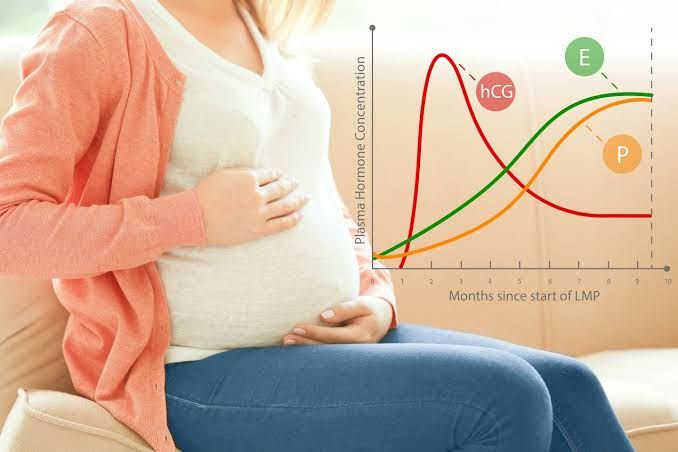
- HCG Secretion Begins: The syncytiotrophoblast cells of the implanted blastocyst begin secreting HCG, signaling pregnancy to the maternal system. HCG levels rise rapidly, doubling approximately every 48-72 hours.
- Corpus Luteum Support: HCG “rescues” the corpus luteum, ensuring continued progesterone production.
3.3. First Trimester (Weeks 1-12): The Period of Organogenesis and Rapid Maternal Adaptation The first trimester is characterized by rapid embryonic development, formation of vital support structures, and significant maternal hormonal shifts.
- Embryonic Development: This is the most critical period for organogenesis, where all major organs and body systems begin to form. The embryo differentiates into distinct germ layers (ectoderm, mesoderm, endoderm), which give rise to all tissues and organs. By the end of this trimester, the embryo is recognizable as a fetus, with rudimentary limbs, a beating heart, and developing brain.
- Placental Development: The placenta, chorion, and amnion develop. The placenta rapidly expands its villi and establishes efficient nutrient and waste exchange pathways with the maternal circulation.
- Hormonal Milieu:
- HCG: Peaks around 8-11 weeks, then declines.
- Progesterone: Steadily rises, primarily from the corpus luteum initially, then increasingly from the placenta. It maintains uterine quiescence and the endometrial lining.
- Estrogen: Gradually rises, initially from the corpus luteum, then from the placenta. Contributes to uterine growth and development of mammary glands.
- Maternal Adaptations:
- Nausea and Vomiting (Morning Sickness): Common, often attributed to rising HCG levels.
- Fatigue: Profound fatigue is common due to high progesterone levels, increased metabolic demands, and emotional adjustments.
- Breast Tenderness and Enlargement: Due to estrogen and progesterone stimulating mammary gland development.
- Frequent Urination: Uterus begins to press on the bladder.
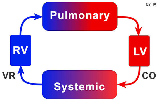
- Increased Blood Volume: Begins to increase, impacting cardiac output.
- Mood Swings: Hormonal fluctuations contribute to emotional lability.
- Cervical Changes: Softening (Goodell’s sign) and bluish discoloration (Chadwick’s sign).
3.4. Second Trimester (Weeks 13-27): Growth, Maturation, and Maternal Comfort Often considered the “golden trimester,” as many early pregnancy discomforts subside, and the mother begins to feel more energetic.
- Fetal Development:
- Rapid growth in length and weight.
- Organ systems continue to mature and specialize (e.g., lungs develop air sacs, brain undergoes significant growth).
- Fetal movements (quickening) are typically felt by 16-20 weeks.
- Sex determination is usually possible via ultrasound.
- Placental Dominance: The placenta fully takes over hormone production (progesterone, estrogen, and others), making the corpus luteum redundant.
- Maternal Adaptations:
- Increased Energy: Nausea subsides, and energy levels often improve.
- Visible Abdomen: The uterus grows significantly, and pregnancy becomes physically apparent.
- Cardiovascular Changes: Blood volume continues to increase (up to 40-50% by term), leading to increased cardiac output. Heart rate may increase by 10-15 bpm.
- Respiratory Changes: Tidal volume increases, leading to a feeling of increased breathlessness, but overall respiratory rate may not change significantly.
- Musculoskeletal Changes: Ligaments soften (due to relaxin), preparing the pelvis for birth, but also contributing to joint laxity and back pain. The center of gravity shifts.
- Skin Changes: Melasma (mask of pregnancy), linea nigra, stretch marks (striae gravidarum) may appear.
- Fetal Movements: A reassuring sign of continued growth and development.
3.5. Third Trimester (Weeks 28-40): Final Preparation and Readiness for Birth The final trimester is marked by significant fetal growth, preparation for labor, and increasing maternal discomfort.
- Fetal Development:
- Rapid weight gain as fat accumulates, preparing for extrauterine life.
- Lungs continue to mature, producing surfactant vital for breathing after birth.
- Brain development accelerates.
- The fetus typically moves into a head-down (cephalic) position in preparation for birth.
- Maternal Adaptations:
- Increased Discomfort: Back pain, pelvic pressure, heartburn (due to uterine pressure on stomach), shortness of breath (uterus presses on diaphragm), leg cramps, edema (swelling) of ankles and feet.
- Braxton Hicks Contractions: Irregular, mild uterine contractions (“practice contractions”) become more frequent.
- Frequent Urination: Due to fetal head engagement and uterine pressure on the bladder.
- Cervical Ripening: The cervix begins to soften, efface (thin), and dilate slightly in preparation for labor, influenced by progesterone withdrawal relative to estrogen.
- “Nesting” Instinct: A surge of energy and a desire to prepare the home for the baby.
- Pre-Labor Signs: Lightening (fetal dropping into the pelvis), increased vaginal discharge (“show”), rupture of membranes (water breaking).
3.6. Labor and Delivery: The culmination of pregnancy, characterized by a complex interplay of hormones, mechanical forces, and maternal effort.
- Hormonal Triggers: A decrease in the progesterone-to-estrogen ratio, increased levels of oxytocin (from the posterior pituitary), and prostaglandins (from the uterus and fetal membranes) initiate labor.
- Uterine Contractions: Coordinated rhythmic contractions of the myometrium, initially mild and infrequent, gradually increasing in intensity, frequency, and duration.
- Stages of Labor:
- First Stage: Cervical effacement and dilation (to 10 cm). This is the longest stage.
- Second Stage: Pushing and delivery of the baby.
- Third Stage: Delivery of the placenta (afterbirth).
- Postpartum: The maternal body begins its recovery, with uterine involution (shrinking back to pre-pregnancy size) and hormonal shifts as the new mother adapts to lactation and non-pregnant physiology.
Conclusion
Pregnancy is a testament to the incredible adaptability and precision of human physiology. Human Chorionic Gonadotropin (HCG) stands as a foundational pillar in this intricate process, indispensable for initiating and sustaining early gestation by rescuing the corpus luteum and ensuring a stable uterine environment. From the moment of conception, through the distinct trimesters of fetal development and maternal adaptation, to the remarkable event of labor and delivery, each physiological event is meticulously coordinated. This comprehensive understanding of HCG’s synthesis and function, coupled with the broader landscape of physiological changes, underscores the remarkable journey that culminates in the miracle of birth.


