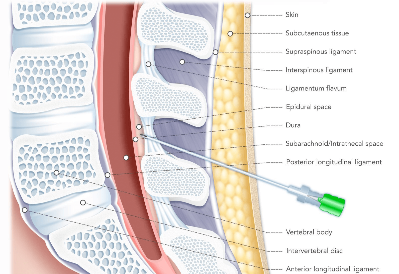
Post-dural puncture headache (PDPH) is a well-recognized iatrogenic complication that can arise following procedures involving the intentional or accidental puncture of the dura mater. Within the field of obstetrics and gynecology, its incidence is particularly relevant due to the high frequency of neuraxial anesthesia (spinal and epidural) administered for labor analgesia, cesarean delivery, and certain gynecological surgeries. While often self-limiting, a severe PDPH can cause significant morbidity, interfering with maternal-infant bonding, breastfeeding, and a patient’s overall recovery.
Understanding the Pathophysiology
The central nervous system is suspended within the protective confines of the dura mater, bathed in cerebrospinal fluid (CSF). This fluid, produced at a rate of approximately 0.35 mL/minute, serves as a buoyant cushion, protecting the brain and spinal cord from trauma. The integrity of the dural sac is crucial for maintaining a stable intracranial pressure.
A PDPH is fundamentally a headache caused by low CSF pressure. When the dura is punctured by a needle, CSF can leak from the subarachnoid space into the epidural space faster than it can be produced. This net loss of CSF volume reduces the buoyant support for the brain. According to the Monro-Kellie doctrine—which posits that the sum of the volumes of brain, CSF, and intracranial blood is constant—the loss of CSF volume leads to two primary consequences that cause pain. First, the brain sags caudally (downward) within the skull, placing traction on pain-sensitive anchoring structures like meninges, cranial nerves, and bridging veins. This traction is exacerbated in an upright position, which explains the hallmark postural nature of the headache. Second, to compensate for the lost CSF volume, a reflexive and compensatory vasodilation of cerebral veins occurs. This increase in blood volume helps to restore total intracranial volume but contributes to a throbbing, vascular-type headache.
Identifying Key Risk Factors
Not every patient who undergoes a dural puncture develops a headache. The risk is influenced by a combination of patient-specific and procedure-related factors.
- Patient-Related Factors:
- Age and Sex: Younger, female patients are at the highest risk. Parturients (women in labor or postpartum) are a particularly vulnerable demographic, possibly due to hormonal changes that affect dural elasticity and the physiological changes of pregnancy.
- Body Mass Index (BMI): Patients with a lower BMI have a higher incidence of PDPH.
- History: A prior history of PDPH or chronic headaches (e.g., migraines) can predispose an individual to developing one.
- Procedure-Related Factors (Largely Modifiable):
- Needle Size (Gauge): This is one of the most significant risk factors. The larger the diameter of the needle, the larger the dural defect and the greater the subsequent CSF leak. For example, an accidental dural puncture (ADP) with a 17-gauge Tuohy epidural needle carries a PDPH risk of over 50%.
- Needle Tip Design: The shape of the needle tip is critical. Traditional “cutting” needles, like the Quincke, have a beveled tip that severs dural fibers, creating a larger, more persistent hole. In contrast, “pencil-point” or non-cutting needles, such as the Sprotte and Whitacre, have a conical tip that separates or spreads the dural fibers. These fibers tend to re-approximate after the needle is withdrawn, significantly reducing the incidence of CSF leakage and PDPH.
- Needle Bevel Orientation: When using a cutting needle for a spinal anesthetic, orienting the bevel parallel to the longitudinal axis of the dural fibers is believed to create a smaller defect, thereby lowering the risk.
- Operator Experience: Less experienced practitioners have a higher rate of multiple attempts and ADP during epidural placement, which directly correlates with an increased risk of PDPH.
Clinical Presentation and Diagnosis
The diagnosis of PDPH is primarily clinical, based on a characteristic history and symptom profile following a known or suspected dural puncture.
- Hallmark Symptom: The defining feature is a postural headache. The pain is typically described as dull, throbbing, or aching, and is most commonly located in the frontal or occipital regions, often radiating to the neck and shoulders. Crucially, the headache worsens significantly within 15-30 minutes of assuming an upright position (sitting or standing) and is dramatically relieved by lying flat (supine). Onset is usually within 24 to 72 hours of the procedure but can be immediate or delayed for several days.
- Associated Symptoms: The traction on cranial nerves and meningeal irritation can produce a constellation of associated symptoms:
- Nausea and Vomiting: Common due to the severity of the pain and autonomic nervous system involvement.
- Auditory Symptoms: Tinnitus (ringing in the ears), hearing loss, or hyperacusis (sensitivity to sound) may occur due to changes in inner-ear fluid pressure.
- Visual Symptoms: Photophobia (light sensitivity) and blurred vision are frequent. Diplopia (double vision), while less common, is a significant finding resulting from the traction and palsy of the abducens nerve (cranial nerve VI).
- Neck Stiffness: Nuchal rigidity can be present, mimicking meningitis.
- Differential Diagnosis: In the postpartum period, it is imperative to rule out other serious causes of headache, including pre-eclampsia/eclampsia, meningitis, cerebral venous thrombosis, subarachnoid hemorrhage, or even a caffeine-withdrawal headache. The postural component is the key differentiator for PDPH, but a thorough neurological exam is essential.
Implementing Management Strategies
Management of PDPH follows a stepwise approach, beginning with conservative measures and escalating to more definitive, invasive treatment if symptoms persist or are severe.
- Conservative Management (First-Line): For mild to moderate headaches, initial management focuses on symptomatic relief and promoting natural healing.
- Supine Positioning: Bed rest and avoiding upright posture minimizes the headache.
- Hydration: Aggressive oral or intravenous fluid administration is often recommended to support CSF production.
- Analgesia: Simple analgesics like acetaminophen and non-steroidal anti-inflammatory drugs (NSAIDs) can help manage the pain. Opioids should be used cautiously.
- Caffeine: Oral or intravenous caffeine (e.g., 300-500 mg) is a cornerstone of conservative therapy. As a potent cerebral vasoconstrictor, it counteracts the painful compensatory vasodilation in the brain.
- Invasive Management: The Epidural Blood Patch (EBP) When conservative measures fail or the headache is debilitating from the outset, the epidural blood patch (EBP) is considered the gold-standard treatment.
- Procedure: Under sterile conditions, approximately 15-20 mL of the patient’s own blood is drawn from a peripheral vein. This autologous blood is then slowly injected into the epidural space at or near the level of the previous dural puncture.
- Mechanism of Action: The EBP works through two proposed mechanisms. First, the injected blood forms a clot or “patch” over the dural defect, mechanically sealing the leak. Second, the volume of blood injected into the epidural space increases subarachnoid pressure (a “tamponade effect”), which immediately elevates CSF pressure, lifts the brain back into its normal position, and relieves the traction-related pain.
- Efficacy and Risks: The EBP has a high success rate, providing immediate and complete relief in 70-98% of patients. A second EBP may be required in some cases. While generally safe, potential risks include back pain at the injection site, transient nerve irritation (radiculopathy), and, very rarely, infection or a repeat dural puncture.
Focusing on Prevention
Given the impact of PDPH, prevention is the most effective strategy. This relies heavily on meticulous technique during neuraxial procedures.
- Use Pencil-Point Needles: For spinal anesthesia, the routine use of small-gauge, non-cutting (Sprotte or Whitacre) needles has been shown to dramatically reduce the incidence of PDPH compared to cutting (Quincke) needles.
- Optimize Technique for Epidurals: When placing an epidural catheter, careful identification of the epidural space is critical to avoid ADP. Using techniques like the loss-of-resistance to saline instead of air may reduce the risk.
- Management of a Known ADP: If an ADP occurs with a large-bore epidural needle, one preventive strategy involves threading the epidural catheter into the intrathecal space and running a very dilute spinal anesthetic. This may “stent” the dural hole and promote an inflammatory healing response, potentially reducing the risk of a subsequent PDPH. The use of a prophylactic EBP immediately after ADP is controversial and not routinely recommended.
In conclusion, post-dural puncture headache remains a significant clinical challenge in obstetrics and gynecology. A thorough understanding of its pathophysiology, risk factors, and classic postural presentation is essential for accurate diagnosis. Management should proceed logically from conservative therapies to the highly effective epidural blood patch. Ultimately, the greatest impact on patient well-being comes from a commitment to preventive strategies, primarily through the selection of appropriate equipment and meticulous procedural technique, ensuring a safer and more comfortable postpartum experience.
References
- Turnbull, D. K., & Shepherd, D. B. (2003). Post-dural puncture headache: pathogenesis, prevention and treatment. British Journal of Anaesthesia, 91(5), 718-729.
- Headache Classification Committee of the International Headache Society (IHS). (2018). The International Classification of Headache Disorders, 3rd edition. Cephalalgia, 38(1), 1-211.
- Arevalo-Rodriguez, I., Ciapponi, A., Roqué i Figuls, M., Muñoz, L., & Bonfill Cosp, X. (2016). Posture and fluids for preventing post-dural puncture headache. Cochrane Database of Systematic Reviews, (3), CD009199.
- Gauthama, P., Chilkoti, G. T., Mohta, M., & Agarwal, D. (2020). Post Dural Puncture Headache in Parturients: A Review of Advancements in the Last Decade. Indian Journal of Anaesthesia, 64(2), 90–98.
- Vallejo, M. C., Mandell, G. L., Sabo, D. P., & Ramanathan, S. (2000). Postdural puncture headache: a randomized comparison of five spinal needles in obstetric patients. Anesthesia & Analgesia, 91(4), 916-920.


