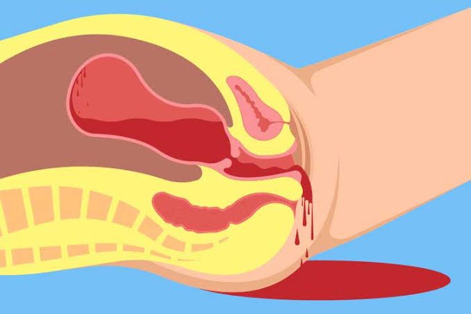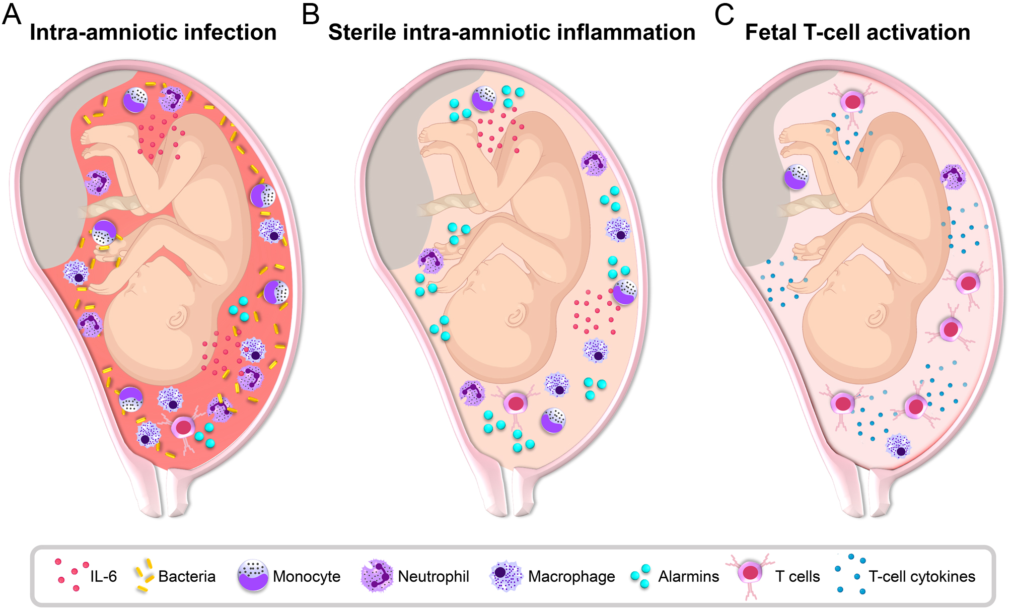
Obstetric haemorrhage remains a leading cause of global maternal mortality, representing a critical emergency that demands a rapid, coordinated, and systematic response from the entire multidisciplinary team. Effective management hinges on anticipation, preparedness, and a clear understanding of the complex protocols designed to control bleeding and resuscitate the patient.
Aetiology: Understanding the Causes
The causes of major obstetric haemorrhage are broadly categorized into antepartum (before delivery) and postpartum (after delivery) events, with the “Four T’s” mnemonic (Tone, Tissue, Trauma, Thrombin) providing a crucial framework for diagnosing postpartum haemorrhage (PPH).
- Tone (Uterine Atony): This is the most common cause of PPH, accounting for approximately 70-80% of cases. It refers to the failure of the uterus to contract effectively after delivery, leading to rapid blood loss from the placental site. Risk factors include uterine overdistension (e.g., macrosomia, multiple gestation, polyhydramnios), prolonged or precipitous labour, chorioamnionitis, and the use of uterine-relaxing drugs.
- Trauma: Injury to the genital tract can occur spontaneously or iatrogenically during delivery. This includes cervical, vaginal, or perineal lacerations, uterine rupture, and an extension of an episiotomy.
- Tissue: Retained products of conception, such as placental fragments or membranes, prevent the uterus from contracting fully. This includes conditions like placenta accreta spectrum (PAS), where the placenta abnormally invades the uterine wall.
- Thrombin (Coagulopathy): Any pre-existing or acquired coagulation disorder can cause or exacerbate haemorrhage. This may be pre-existing (e.g., von Willebrand disease) or acquired, such as disseminated intravascular coagulation (DIC) secondary to placental abruption, amniotic fluid embolism, or severe pre-eclampsia.
Symptoms and Signs: Early Recognition is Key
Recognition must be swift. Signs extend beyond simple observed blood loss, as underestimation is common, and physiological changes in pregnancy can mask hypovolaemia until sudden decompensation occurs.
- Obvious Symptoms: Visual estimation of heavy vaginal bleeding (though often underestimated), or the presence of concealed bleeding (e.g., intra-abdominal or broad ligament haematoma).
- Signs of Hypovolaemic Shock: Tachycardia (often the earliest sign), tachypnoea, hypotension, pallor, cool and clammy skin, decreased capillary refill, oliguria, and altered mental status (agitation, confusion, or drowsiness).
- Specific Signs: A soft, “boggy” uterus palpable above the umbilicus suggests uterine atony. Visualisation of a laceration or continuous bleeding despite a firm uterus points to trauma.
Management of Unanticipated Haemorrhage: The Systematic Approach
The management of major haemorrhage is simultaneous and multimodal, following a standardized protocol like the one advocated by the Royal College of Obstetricians and Gynaecologists (RCOG).
- Call for Help: Immediately activate the major haemorrhage protocol, summoning senior obstetricians, anaesthetists, midwives, and haematologists.
- Resuscitation (ABC Approach):
- Airway & Breathing: Administer high-flow oxygen via a non-rebreather mask. Secure the airway early if consciousness is impaired.
- Circulation: Obtain large-bore intravenous access (x2 cannulae, 14-16G). Commence rapid infusion of warmed crystalloid solutions (e.g., Hartmann’s solution) followed immediately by blood products as per protocol.
- Monitor & Investigate: Continuously monitor vital signs. Send urgent blood samples for full blood count (FBC), coagulation screen (including fibrinogen), and crossmatch (4-6 units minimum). Point-of-care testing, such as viscoelastic haemostatic assays (TEG/ROTEM), can provide rapid real-time guidance on coagulation status.
- Identify and Treat the Cause: While resuscitation is ongoing, the primary obstetric cause must be identified and treated.
Anaesthetic Management
The anaesthetist plays a pivotal role in resuscitation, analgesia, and preparation for surgical intervention. For an unstable patient, rapid sequence induction (RSI) and general anaesthesia are typically preferred to secure the airway and control ventilation. For a stable patient with a predicted difficult airway, neuraxial anaesthesia (e.g., an existing epidural) may be extended. The anaesthetist manages fluid resuscitation, administers blood products and drugs, and provides haemodynamic support with vasopressors as needed.
Uterotonic Drugs: First-Line Medical Management
These are the first-line pharmacological agents used to induce uterine contraction and combat atony, often administered in sequence:
- Oxytocin: First-line agent, given as an IV bolus (typically 5IU) followed by a continuous infusion (e.g., 40IU in 500ml saline over 4 hours).
- Ergometrine: Causes powerful tetanic uterine contractions. Often combined with oxytocin as Syntometrine®. Contraindicated in hypertension and pre-eclampsia.
- Carboprost (Hemabate®): A prostaglandin F2-alpha analogue, given intramyometrially. Effective in many cases refractory to oxytocin/ergometrine. Contraindicated in asthma.
- Misoprostol: A prostaglandin E1 analogue, used sublingually or rectally. Particularly useful in low-resource settings due to its stability.
Surgical Management
If medical management fails, prompt escalation to surgical intervention is critical.
- Examination under Anaesthesia (EUA): To identify and repair traumatic causes.
- Uterine Compression Sutures: The B-Lynch suture is a well-known technique that mechanically compresses the uterus.
- Vessel Ligation: Stepwise ligation of the uterine, ovarian, or internal iliac arteries can be performed to reduce pulse pressure and blood flow.
- Peripartum Hysterectomy: The definitive life-saving surgical procedure for uncontrolled haemorrhage, performed when all other measures have failed. This is a radical but necessary step to save the mother’s life.
Radiological Management
In appropriate, stable, or anticipated cases (e.g., known placenta accreta), interventional radiology offers minimally invasive options.
- Uterine Artery Embolisation (UAE): A catheter is guided into the uterine arteries, and embolic material (e.g., gel foam particles) is injected to block blood flow and stop the bleeding. This requires a stable patient and available expertise.
Transfusion Practice & Haemostatic Support
Modern transfusion in major obstetric haemorrhage follows a massive transfusion protocol (MTP), which provides balanced ratios of blood components to mimic whole blood and correct coagulopathy proactively.
- Packed Red Blood Cells (PRBCs): To restore oxygen-carrying capacity.
- Fresh Frozen Plasma (FFP): Provides clotting factors.
- Platelets: Transfused to maintain a count >75 x 10⁹/L.
- Cryoprecipitate or Fibrinogen Concentrate: Critical for correcting hypofibrinogenaemia, a key feature of obstetric coagulopathy. Aim to maintain fibrinogen >2.0 g/L.
- Tranexamic Acid: An antifibrinolytic agent shown to reduce mortality in bleeding postpartum women when given within 3 hours of birth (as per the WOMAN trial). A standard dose is 1g IV administered slowly.
Intraoperative Cell Salvage (ICS)
ICS is now recognised as a safe and valuable technique in obstetrics. It involves collecting, washing, and filtering the patient’s own lost blood before re-infusing it. It reduces the need for allogenic blood transfusion and is considered safe in RhD-negative women with appropriate anti-D immunoglobulin administration.
Additional Drugs
Beyond uterotonics and tranexamic acid, other agents may be used:
- Recombinant Factor VIIa (rFVIIa): A powerful haemostatic agent used as a last resort in catastrophic, uncontrollable bleeding despite conventional treatment. Its use is off-label and requires expert haematological guidance due to thrombotic risks.
- Calcium: Ionised calcium levels often drop during massive transfusion (due to citrate in blood products) and impair cardiac function and coagulation. Regular monitoring and replacement are essential.
Subsequent Management
Surviving the initial event is followed by a critical recovery phase.
- High Dependency Unit (HDU)/ICU Admission: For ongoing close monitoring of haemodynamics, fluid balance, and laboratory parameters.
- Continued Vigilance for Complications: Including ongoing oozing due to coagulopathy, transfusion-related reactions (e.g., TRALI, TACO), electrolyte imbalances, and sepsis.
- Psychological Support: A traumatic near-miss event can have profound psychological impacts on the mother and her family, necessitating debriefing and mental health support.
- Debrief and Documentation: A full team debrief is essential for clinical governance, learning, and providing a clear account for the patient’s notes and future pregnancies.
In conclusion, the successful management of obstetric haemorrhage relies on a standardized, team-based, and rapid protocol that integrates simultaneous resuscitation, identification of the cause, and a escalating cascade of medical, surgical, and radiological interventions. Preparedness through regular drills and clear hospital protocols is the cornerstone of saving lives.
References
- Royal College of Obstetricians and Gynaecologists (RCOG). (2022). Postpartum Haemorrhage, Prevention and Management (Green-top Guideline No. 52). Retrieved from https://www.rcog.org.uk/guidance/browse-all-guidance/green-top-guidelines/postpartum-haemorrhage-prevention-and-management-green-top-guideline-no-52/
- Mavrides, E., Allard, S., Chandraharan, E., et al. (2016). on behalf of the Royal College of Obstetricians and Gynaecologists. Prevention and Management of Postpartum Haemorrhage. BJOG, 124: e106–e149.
- WOMAN Trial Collaborators. (2017). Effect of early tranexamic acid administration on mortality, hysterectomy, and other morbidities in women with post-partum haemorrhage (WOMAN): an international, randomised, double-blind, placebo-controlled trial. The Lancet, 389(10084), 2105-2116.
- The Association of Anaesthetists of Great Britain and Ireland (AAGBI). (2015). Blood transfusion and the anaesthetist: management of massive haemorrhage. Anaesthesia, 70: 75–83.
- Royal College of Anaesthetists (RCoA). (2021). Guidelines for the Provision of Anaesthesia Services (GPAS) Chapter 9: Guidelines for the Provision of Anaesthetic Services in the Non-Theatre Environment.


