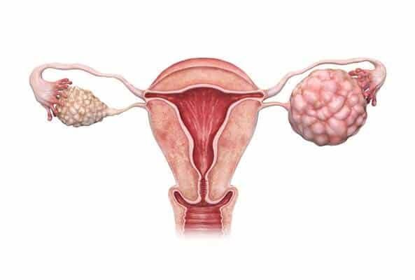
Ovarian neoplasms represent a diverse group of growths arising from the ovary, ranging from functionally benign cysts to aggressive malignancies. Understanding their characteristics, initial management, risk factors, symptoms, and histological classifications is paramount for effective patient care, especially within a framework of value-based healthcare.
Initial Management of a Patient with an Adnexal Mass, with Consideration of Value-Based Care
The discovery of an adnexal mass often presents a clinical challenge, requiring a systematic and patient-centered approach. Initial management prioritizes accurate risk stratification to differentiate between benign and malignant conditions, while simultaneously adhering to principles of value-based care—optimizing outcomes per unit of cost, enhancing patient experience, and reducing unnecessary interventions.
The first step involves a comprehensive history, including age, menopausal status, parity, personal or family history of cancer (especially gynecological or breast), and symptom assessment. A thorough physical examination, including bimanual pelvic and rectovaginal examination, is crucial to assess the mass’s size, mobility, consistency, and any associated tenderness or nodularity, as well as to detect signs of ascites or lymphadenopathy.
Imaging is fundamental, with transvaginal ultrasound (TVS) being the cornerstone. TVS offers detailed visualization of the mass’s morphology, including size, internal septations, solid components, papillary projections, and presence of ascites. Characteristics such as unilocular, anechoic, thin-walled, and smaller lesions are typically indicative of benignity, while multilocular, solid, complex, or rapidly growing masses with papillary structures or increased vascularity raise suspicion for malignancy (International Ovarian Tumor Analysis – IOTA group criteria). Magnetic resonance imaging (MRI) or computed tomography (CT) may be used as secondary imaging modalities for further characterization, surgical planning, or staging, especially for suspicious lesions.
Serum tumor markers, notably CA-125, are often used in conjunction with imaging. While CA-125 is elevated in about 80% of epithelial ovarian cancers, it is not specific and can be raised in benign conditions like endometriosis, uterine fibroids, and pelvic inflammatory disease, particularly in premenopausal women. Therefore, its utility is most significant in postmenopausal women with a suspicious adnexal mass, or for monitoring known malignancy. Other markers like HE4, AFP, hCG, and LDH may be considered depending on the patient’s age and suspected tumor type (e.g., germ cell tumors). Risk of Malignancy Index (RMI) or ADNEX model incorporate multiple parameters (menopausal status, CA-125, ultrasound features) to provide a weighted risk score, guiding referral to gynecologic oncology.
Value-based care dictates a judicious approach. For low-risk, asymptomatic, simple cysts (typically <5-7 cm in premenopausal women), conservative management with watchful waiting and follow-up ultrasound is often appropriate, avoiding unnecessary surgery or invasive procedures. This minimizes patient burden, reduces complications, and controls healthcare costs. For higher-risk or symptomatic lesions, timely referral to a gynecologic oncologist is critical. Shared decision-making, involving a thorough discussion of risks, benefits, and alternatives with the patient, is paramount, ensuring treatment aligns with patient values and preferences. Over-investigation or prophylactic surgery for low-risk findings is discouraged, while under-investigation of high-risk findings could delay life-saving treatment.
Comparing the Characteristics of Functional Cysts, Benign Ovarian Neoplasms, and Ovarian Cancers
Distinguishing these three categories is crucial for appropriate management. They differ significantly in their etiology, natural history, imaging characteristics, and clinical implications.
- Functional Cysts: These are the most common type of adnexal mass, arising from the normal physiological process of ovulation. They are non-neoplastic and typically resolve spontaneously within 1-3 menstrual cycles.
- Examples: Follicular cysts (failed follicle rupture), Corpus luteum cysts (persistent corpus luteum).
- Characteristics: Usually unilocular, anechoic (fluid-filled), thin-walled, smooth interior, and typically less than 10 cm in diameter on ultrasound. They are generally asymptomatic but can cause pelvic pain if they rupture or torse. CA-125 levels are usually normal.
- Benign Ovarian Neoplasms: These are true tumors, meaning they involve abnormal cell growth, but they do not invade surrounding tissues or metastasize. They have growth potential but are not life-threatening unless they cause complications like torsion or rupture.
- Examples:
- Epithelial: Serous cystadenoma, Mucinous cystadenoma, Endometrioid adenofibroma, Brenner tumor.
- Germ Cell: Mature cystic teratoma (dermoid cyst) – containing diverse tissues like hair, teeth, fat.
- Sex Cord-Stromal: Fibroma, Thecoma.
- Characteristics: Often persist or grow larger than functional cysts, and may have more complex imaging features (e.g., septations, solid components, calcifications in dermoids). However, they typically lack features highly suspicious for malignancy like thick irregular septations, large solid components with internal vascularity, or ascites. CA-125 may be mildly elevated in some benign conditions (e.g., endometriosis, fibroma), but not typically to the levels seen in malignancy.
- Examples:
- Ovarian Cancers (Malignant Ovarian Neoplasms): These are characterized by uncontrolled cell growth, invasion of adjacent tissues, and the potential to metastasize to distant sites. They represent a significant health challenge due to their often late presentation.
- Examples: High-grade serous carcinoma (most common), Mucinous carcinoma, Endometrioid carcinoma, Clear cell carcinoma (all epithelial); Dysgerminoma, Endodermal sinus tumor (germ cell); Granulosa cell tumor, Sertoli-Leydig cell tumor (sex cord-stromal).
- Characteristics: Imaging typically reveals complex masses with solid components, thick or irregular septations, papillary projections, ascites, and often evidence of increased blood flow (Doppler). Rapid growth is also a concerning sign. CA-125 levels are often significantly elevated, especially in epithelial ovarian cancer, and may be accompanied by other tumor markers depending on the histological type. Clinically, symptoms are often persistent and progressive.
Risk Factors and Protective Factors for Ovarian Cancer, with Consideration of Population Health Implications and the Impact of Social and Environmental Factors
Understanding the factors influencing ovarian cancer risk is crucial for prevention strategies, targeted screening in high-risk groups, and public health initiatives.
- Risk Factors:
- Genetic Predisposition: The strongest risk factor. Mutations in BRCA1 and BRCA2 genes significantly increase risk (up to 40-60% lifetime risk). Other hereditary cancer syndromes like Lynch syndrome (hereditary non-polyposis colorectal cancer, HNPCC) also confer increased risk. Family history of ovarian, breast, or colorectal cancer is a significant red flag.
- Reproductive Factors: Nulliparity (never having children), infertility (regardless of cause), early menarche (<12 years), late menopause (>55 years), and endometriosis increase the lifetime number of ovulatory cycles, which some theories link to increased epithelial damage and repair.
- Age: Risk increases with age, with most diagnoses occurring after menopause.
- Obesity: Linked to increased chronic inflammation and altered hormone metabolism, contributing to higher risk.
- Hormone Replacement Therapy (HRT): Combined estrogen-progestin HRT, particularly long-term use, has been associated with a small increased risk.
- Talc Use: While controversial, some studies suggest a possible link between perineal talc use and epithelial ovarian cancer.
- Population Health Implications: Genetic counseling and testing for high-risk individuals are vital. Public health campaigns promoting healthy lifestyle choices (diet, exercise) to combat obesity are relevant for general cancer prevention. Access to reproductive healthcare and family planning can also influence parity trends.
- Protective Factors:
- Oral Contraceptive Pills (OCPs): Use of OCPs for 5 years or more can reduce risk by up to 50%, with protection persisting for decades after cessation. This is thought to be due to the suppression of ovulation.
- Multiparity: Each full-term pregnancy further reduces risk, likely due to prolonged periods of anovulation.
- Breastfeeding: Also associated with a reduced risk.
- Tubal Ligation, Hysterectomy: These procedures have been shown to reduce ovarian cancer risk, possibly by preventing carcinogenic agents from reaching the ovaries or by removing potential sites of origin for some serous cancers (e.g., from the fallopian tube fimbriae).
- Risk-Reducing Salpingo-Oophorectomy (RRSO): For women with BRCA1/2 mutations, surgical removal of ovaries and fallopian tubes significantly reduces ovarian and fallopian tube cancer risk.
- Population Health Implications: Public health education on the protective effects of OCPs and breastfeeding can empower women to make informed choices. Access to family planning services and options for elective surgical procedures (like tubal ligation during C-sections) also play a role.
- Impact of Social and Environmental Factors:
- Socioeconomic Status (SES): Lower SES can indirectly increase risk through disparities in access to healthcare, preventative education, and genetic counseling. It can also influence lifestyle factors such as diet, exercise, and exposure to environmental stressors.
- Environmental Toxins: While direct links to ovarian cancer are less established compared to other cancers, the general principle of environmental carcinogen exposure cannot be entirely discounted. Further research is needed.
- Healthcare Access & Disparities: Limited access to gynecological care, especially for routine check-ups and symptom evaluation, can lead to delayed diagnosis, particularly in underserved populations. This contributes to disparities in ovarian cancer outcomes. Social support networks and cultural understanding also influence health-seeking behaviors.
Symptoms and Physical Findings Associated with Ovarian Cancer
Ovarian cancer is often referred to as a “silent killer” because its early symptoms are typically vague, non-specific, and easily attributable to more common, benign conditions. This often leads to a late diagnosis when the disease has already spread.
- Symptoms (Often Persistent and Progressive):
- Bloating: Persistent abdominal swelling or distension, not related to diet.
- Pelvic or Abdominal Pain: Persistent discomfort, pressure, or cramping in the lower abdomen or pelvis.
- Difficulty Eating or Feeling Full Quickly (Early Satiety): Feeling full after eating only a small amount of food.
- Urinary Symptoms: Increased frequency or urgency of urination.
- Other Non-Specific Symptoms: Fatigue, indigestion, heartburn, nausea, changes in bowel habits (constipation or diarrhea), unexplained weight gain or loss, dyspareunia (painful intercourse), and irregular bleeding (less common but can occur).
The critical distinction is the persistence and progression of these symptoms, occurring almost daily for several weeks, unlike transient indigestion or menstrual bloating. Women, especially those over 50 or with risk factors, should be advised to seek medical attention for new, persistent, and recurrent symptoms.
- Physical Findings (Often Indicate Advanced Disease):
- Abdominal Distension: Due to ascites (fluid accumulation in the abdomen) or a large tumor mass.
- Palpable Pelvic Mass: On bimanual examination, the mass may be fixed, irregular, nodular, or firm.
- Nodularity in the Cul-de-Sac/Pouch of Douglas: Detected on rectovaginal examination, indicating peritoneal seeding.
- Pleural Effusion: Fluid in the chest cavity, often on the right side, suggesting diaphragmatic involvement.
- Advanced Metastatic Signs:
- Sister Mary Joseph Nodule: A palpable umbilical nodule indicative of peritoneal metastasis.
- Virchow’s Node: Enlarged, hard, non-tender supraclavicular lymph node, indicating lymphatic spread.
- Leg Swelling: Due to lymphatic or venous obstruction.
- Cachexia: General wasting and malnutrition in advanced stages.
The Three Histological Categories of Ovarian Neoplasms
Ovarian neoplasms are broadly classified into three main histological categories based on their cell of origin. This classification is critical because each category has distinct epidemiology, clinical behavior, and treatment responsiveness.
- a) Epithelial Tumors:
- Origin: Derived from the ovarian surface epithelium (or Müllerian remnants, or via migration from the fallopian tube fimbriae, particularly for high-grade serous cancer). They account for approximately 90% of all ovarian cancers and a significant proportion of benign ovarian neoplasms.
- Subtypes (Malignant Examples):
- Serous Carcinoma: The most common type of epithelial ovarian cancer (50-70%), often bilateral. High-grade serous carcinoma (HGSC) accounts for the majority of deaths and is frequently aggressive, often originating in the fallopian tube fimbriae (STICS – serous tubal intraepithelial carcinoma). Low-grade serous carcinoma is rarer and less aggressive.
- Mucinous Carcinoma: Accounts for 5-10% of ovarian cancers, often very large and multiloculated. Can be difficult to distinguish from gastrointestinal primary cancers metastasizing to the ovary (Krukenberg tumors).
- Endometrioid Carcinoma: Accounts for ~10% of ovarian cancers and is often associated with endometriosis or endometrial carcinoma.
- Clear Cell Carcinoma: Accounts for ~5-10% of ovarian cancers, also strongly associated with endometriosis. Tends to be more aggressive with a poorer prognosis if not diagnosed early.
- Brenner Tumor: Most are benign, but a small percentage can be borderline or malignant. Characterized by transitional cell-like epithelium.
- Undifferentiated Carcinoma: Lacks specific differentiation and is usually aggressive.
- Clinical Features: Typically affect older women (postmenopausal). CA-125 is a common tumor marker.
- b) Germ Cell Tumors:
- Origin: Arise from the primordial germ cells within the ovary. These tumors are relatively rare, accounting for less than 5% of ovarian cancers.
- Subtypes (Malignant Examples):
- Dysgerminoma: The most common malignant germ cell tumor (30-40% of germ cell cancers). It is sensitive to radiation and chemotherapy. It’s the ovarian counterpart of testicular seminoma.
- Endodermal Sinus Tumor (Yolk Sac Tumor): Highly aggressive, characterized by elevated serum alpha-fetoprotein (AFP).
- Embryonal Carcinoma: Highly malignant, often mixed with other germ cell elements.
- Choriocarcinoma: Very rare, highly aggressive, characterized by elevated serum human chorionic gonadotropin (hCG).
- Teratoma:
- Mature Cystic Teratoma (Dermoid Cyst): The most common germ cell tumor (benign), containing mature tissues from all three germ layers (e.g., skin, hair, teeth, bone, fat).
- Immature Teratoma: Malignant, containing immature neural or other embryonic tissues.
- Clinical Features: Primarily affect children and young women. Specific tumor markers (AFP, hCG, LDH) are often elevated and useful for diagnosis and monitoring. Highly curable even in advanced stages with platinum-based chemotherapy.
- c) Sex Cord-Stromal Tumors:
- Origin: Derived from the specialized stromal cells of the ovary (e.g., granulosa, theca, Sertoli, Leydig cells). These tumors account for approximately 5-10% of ovarian neoplasms, with most being benign.
- Subtypes (Examples):
- Granulosa Cell Tumor: The most common malignant sex cord-stromal tumor. Often estrogen-producing, leading to symptoms like abnormal uterine bleeding (postmenopausal) or precocious puberty (prepubertal girls). Inhibin B is a useful tumor marker.
- Sertoli-Leydig Cell Tumor: Often androgen-producing, leading to virilization (e.g., hirsutism, voice deepening).
- Fibroma/Thecoma: Typically benign, non-functional tumors of the ovarian stroma. Fibromas are often associated with Meigs’ syndrome (fibroma, ascites, right-sided pleural effusion).
- Clinical Features: Can occur at any age. Known for their hormonal activity, which can lead to specific endocrine manifestations. They can be unilateral and are often slow-growing.
In conclusion, the complexity of ovarian neoplasms demands a nuanced and evidence-based approach. From the initial evaluation of an adnexal mass with value-based considerations to understanding the distinct characteristics of functional cysts, benign neoplasms, and cancers, and appreciating the multifaceted nature of risk factors and symptoms, comprehensive knowledge is essential. Furthermore, the precise histological classification guides prognosis and dictates appropriate therapeutic strategies. Continual education, vigilance for subtle symptoms, and adherence to established guidelines are crucial for improving outcomes for women affected by ovarian neoplasms.
References
- ACOG Practice Bulletin No. 174: Evaluation and Management of Adnexal Masses. (2016). Obstetrics & Gynecology, 128(5), e210–e226.
- American Cancer Society. (2023). Ovarian Cancer Risk Factors. Retrieved from https://www.cancer.org/cancer/types/ovarian-cancer/causes-risks-prevention/risk-factors.html
- Bowman, M., & Kumar, A. (2023). Adnexal Mass. In: StatPearls. Treasure Island (FL): StatPearls Publishing.
- Goff, B. A., Mandel, L. S., Muntz, H. G., & Fuller, A. F. (2000). Ovarian cancer diagnosis. Cancer, 89(10), 2095-2104.
- International Ovarian Tumor Analysis (IOTA) group. (2023). Risk models for adnexal masses. Retrieved from https://www.iotagroup.org/
- Kurman, R. J., Carcangiu, L. M., Herrington, C. S., & Young, R. H. (Eds.). (2014). WHO Classification of Tumours of Female Reproductive Organs. (4th ed.). Lyon: International Agency for Research on Cancer.
- Peres, L. C., Cushing-Haugen, K. L., Köbel, M., Harris, H. R., & Bandera, E. V. (2020). The Etiology of Ovarian Cancer: A Historical Perspective and a Molecular In-Depth Analysis. Cancer Research, 80(13), 2680-2691.
- Siegel, R. L., Miller, K. D., & Jemal, A. (2020). Cancer statistics, 2020. CA: A Cancer Journal for Clinicians, 70(1), 7-30.


