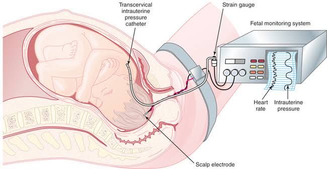
Intrapartum fetal surveillance (IFS) constitutes a critical component of obstetric care, aiming to monitor the fetal physiological response to the profound stresses of labor. The primary goal of IFS is the early detection of fetal compromise, particularly hypoxia or acidosis, which, if unaddressed, can lead to permanent neurological injury or fetal death. Effective surveillance requires not only technological competence but also refined interpretive skill, integrated teamwork, and adherence to principles of value-based care.
Explaining Intrapartum Fetal Surveillance
Intrapartum Fetal Surveillance is the continuous or intermittent assessment of fetal well-being during active labor and delivery. During uterine contractions, blood flow to the placenta is intermittently reduced, creating periods of potential stress for the fetus. Most fetuses tolerate these transient hypoxic episodes well. However, those with pre-existing conditions (such as placental dysfunction, growth restriction, or maternal chronic illnesses) may decompensate.
The core objectives of IFS are:
- Early Identification: To recognize patterns on the fetal monitor that signify progressive hypoxemia or acidosis before the onset of irreversible injury.
- Timely Intervention: To guide necessary interventions, ranging from simple intrauterine resuscitation measures to urgent operative delivery.
- Preventing Unnecessary Intervention: To differentiate benign or compensatory fetal responses from true pathological patterns, thereby reducing the rate of unnecessary cesarean sections or instrumental deliveries.
A robust understanding of fetal physiology—including mechanisms of oxygen regulation, cardiac output distribution, and acid-base balance—is essential for accurately evaluating surveillance findings.
Description of the Methods of Fetal Surveillance
Fetal surveillance methods are broadly categorized into primary, non-invasive techniques and secondary, invasive assessment tools.
1. Intermittent Auscultation (IA)
For women classified as low-risk (no significant maternal or fetal co-morbidities), intermittent auscultation remains the recommended surveillance method by many professional bodies, particularly due to its association with lower rates of obstetric intervention without increasing the risk of adverse neonatal outcomes.
- Methodology: IA involves listening to the fetal heart rate (FHR) using a Doppler ultrasound device or fetoscope for a specified duration (e.g., 60 seconds) following a contraction.
- Frequency: Standards typically require auscultation every 15–30 minutes in the active phase of the first stage of labor and every 5–15 minutes in the second stage.
- Assessment: The clinician primarily assesses the FHR baseline and the presence of accelerations or decelerations immediately following the contraction.
2. Continuous Electronic Fetal Monitoring (EFM)
EFM is the standard of care for high-risk pregnancies and is widely utilized in most labor and delivery units globally. EFM provides a continuous, graphic record of the FHR and uterine activity (UA).
- External Monitoring (Non-Invasive):
- Tocodynamometer (Toco): Measures the frequency and duration of contractions via a belt placed over the maternal abdomen. It does not accurately measure intensity.
- External Doppler Transducer: Uses ultrasound to detect the fetal heart movement and calculate the FHR.
- Internal Monitoring (Invasive): Used when external signals are inadequate or when precise measurements are crucial.
- Fetal Scalp Electrode (FSE): Attached directly to the fetal scalp to provide a highly accurate QRS complex count, ensuring true beat-to-beat variability assessment. Requires ruptured membranes.
- Intrauterine Pressure Catheter (IUPC): Placed inside the uterus to accurately measure contraction intensity (in mmHg), resting tone, and Montevideo Units (MVUs). Requires ruptured membranes.
3. Adjunctive and Secondary Methods
When EFM tracings are concerning (Category II patterns), additional tests may be employed to assess current fetal acid-base status:
- Fetal Scalp Stimulation or Vibroacoustic Stimulation (VAS): A non-invasive test used to elicit an FHR acceleration. The presence of an acceleration (reactive fetus) strongly suggests a normal fetal pH.
- Fetal Scalp Blood Sampling (FSBS): An invasive procedure measuring fetal blood pH or lactate levels. This is less common today but serves as the historical gold standard to confirm acidosis associated with pathological FHR patterns.
- Amnioinfusion: The introduction of warmed sterile saline into the uterus via an IUPC. Primarily used to alleviate severe or repetitive variable decelerations caused by umbilical cord compression.
Interpreting Intrapartum Electronic Fetal Heart Rate Monitoring
Systematic interpretation is essential for clinical decision-making. The National Institute of Child Health and Human Development (NICHD) and the American College of Obstetricians and Gynecologists (ACOG) established a standardized vocabulary for FHR interpretation, based on four primary components and a three-tiered classification system.
A. Components of FHR Interpretation
- Baseline FHR: The approximate mean FHR rounded to increments of 5 beats per minute (bpm) during a 10-minute segment, excluding accelerations and decelerations. Normal range is 110–160 bpm.
- Tachycardia: Baseline >160 bpm (often related to fever, infection, or maternal medication).
- Bradycardia: Baseline <110 bpm (may indicate late fetal hypoxia or congenital cardiac issues).
- Baseline FHR Variability: Fluctuations in the baseline FHR that are irregular in amplitude and frequency. This is the single most reliable indicator of fetal oxygenation and neurological health.
- Absent: Amplitude range undetectable (ominous).
- Minimal: Amplitude range detectable but 5 bpm or less (concerning).
- Moderate: Amplitude range 6–25 bpm (indicates a well-oxygenated fetus).
- Marked: Amplitude range >25 bpm (uncertain significance, potentially associated with early hypoxia or fetal movement).
- Accelerations: Abrupt increases in FHR above the baseline.
- Prior to 32 weeks gestation: Peak must be 10 bpm above baseline lasting 10 seconds (10×10).
- At or after 32 weeks gestation: Peak must be 15 bpm above baseline lasting 15 seconds (15×15).
- Significance: Accelerations are reassuring and virtually exclude acute fetal academia.
- Decelerations: Decreases in FHR below the baseline, categorized by their timing relative to uterine contractions (UC):
- Early Decelerations: Symmetrical, gradual decrease and return of FHR, mirroring the UC. Nadir (lowest point) of the decel occurs at the peak of the contraction. Cause: Head compression. Significance: Benign.
- Variable Decelerations: Abrupt, jagged decreases in FHR varying widely in shape, depth, and duration. Cause: Umbilical cord compression. Significance: Mild to moderate are common; recurrent, severe, or slow-to-recover variables may indicate escalating hypoxemia.
- Late Decelerations: Symmetrical, gradual decrease and return of FHR, occurring after the peak of the contraction. Cause: Uteroplacental insufficiency (a drop in oxygen supply). Significance: Non-reassuring, often associated with fetal hypoxia and acidosis.
- Prolonged Decelerations: Decelerations lasting 2 minutes or more but less than 10 minutes (over 10 minutes constitutes a baseline change). Cause: Acute, transient events (e.g., hypotension, prolonged cord compression). Significance: Requires urgent intervention.
B. The Three-Tier FHR Interpretation System
The NICHD/ACOG system classifies FHR tracings into three categories, guiding immediate clinical action:
| Category | Definition | Clinical Implication | Management |
|---|---|---|---|
| Category I (Normal) | Baseline 110–160 bpm, moderate variability, accelerations present or absent, no late or variable decelerations, early decelerations present or absent. | Strongly predictive of normal fetal acid-base status. | Requires routine fetal monitoring. |
| Category III (Abnormal) | Predictive of abnormal fetal acid-base status. Includes: Absent variability and any of the following: Recurrent late decelerations, Recurrent variable decelerations, or Bradycardia. OR Sinusoidal pattern. | Requires immediate intervention (intrauterine resuscitation) and consideration for urgent delivery if patterns persist despite efforts. | |
| Category II (Indeterminate) | All tracings not classified as Category I or III. Includes minimal or marked variability, absent accelerations after stimulation, episodic or recurrent variable decelerations, late decelerations without absent variability, or tachycardia. | Requires ongoing evaluation, surveillance, and intrauterine resuscitation effort. Represents the majority of tracings. | Aggressive intrauterine resuscitation (IV fluids, position change, oxygen, stopping oxytocin); continuous reassessment; consideration of secondary tests (e.g., VAS). |
Intraprofessional Team, Value-Based Care, and Patient Safety
Interpretation of EFM is not a solitary task; it is a dynamic process involving a coordinated interprofessional team committed to applying principles of value-based care and maximizing patient safety.
A. The Role of the Interprofessional Team
Optimal IFS hinges on effective communication and defined roles:
- The Labor and Delivery Nurse: The nurse provides continuous, minute-by-minute EFM surveillance. They are often the first to recognize deviations and initiate immediate intrauterine resuscitation measures (e.g., repositioning the mother, increasing IV fluids, administering tocolytics). The nurse utilizes standardized communication tools (like SBAR) to effectively escalate concerns to the provider.
- The Obstetric Provider (Physician/Midwife): The provider integrates the EFM interpretation with the mother’s clinical context (labor stage, potential etiology, maternal status) to create a management plan. They must be readily available to perform interventions (e.g., FSE placement, operative delivery).
- Anesthesia and Neonatal Teams: These teams must be alerted proactively if a Category II tracing fails to resolve or if a Category III tracing develops, ensuring a rapid response for urgent delivery and immediate neonatal stabilization.
B. Consideration of Value-Based Care
Value-based care (VBC) in obstetrics dictates that care delivered provides the highest quality outcome at the lowest possible cost, shifting the focus from volume (number of C-sections) to value (healthy mother and baby).
- Minimizing Unnecessary Intervention: Over-interpretation of Category II tracings often leads to unnecessary operative delivery. VBC promotes the use of IA for low-risk women and emphasizes secondary tests (VAS, scalp pH) to confirm fetal well-being before proceeding to C-section. This reduces maternal morbidity, shortens hospital stays, and lowers lifetime healthcare costs associated with operative delivery.
- Standardization of Practice: Clear, evidence-based protocols prevent clinicians from relying on subjective judgment, which reduces variability in care. Using the standardized NICHD/ACOG terminology decreases miscommunication and improves efficiency.
- Preventing Adverse Outcomes: While avoiding unnecessary intervention is key, the highest value in IFS is the prevention of severe hypoxic-ischemic encephalopathy (HIE). Although rare, HIE results in catastrophic long-term costs. Effective surveillance, timely recognition of Category III patterns, and rapid response represent the essential high-value steps to avoid this outcome.
C. Impact on Patient Safety
Patient safety is paramount and is enhanced through structured vigilance and education:
- Simulation and Drills: Regular training, including fetal compromise simulation drills, ensures that the entire interprofessional team can recognize, communicate, and execute an emergency response (e.g., C-section readiness) within established time benchmarks (often 30 minutes from decision to incision).
- System Redundancy: Establishing protocols for mandatory second opinions for persistent Category II or all Category III tracings ensures that no single clinician operates in isolation and reduces the likelihood of interpretive error.
- Documentation: Robust and accurate documentation, including maternal actions taken (IU resuscitation measures) and the FHR response, provides a continuous record of the clinical trajectory and supports future quality improvement initiatives.
In conclusion, effective intrapartum fetal surveillance is a sophisticated clinical skill demanding continuous education, standardized terminology, and seamless collaboration. By integrating reliable methods of monitoring with disciplined interpretation and a team-based approach rooted in value-based care, clinicians can maximize the chances of a healthy outcome for both mother and infant while minimizing unwarranted obstetric intervention.


