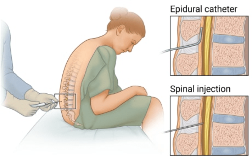
Epidural analgesia is a cornerstone of modern pain management, offering effective relief for a wide range of conditions, from labor pain and postoperative pain to chronic pain management. The efficacy and safety of this technique rely heavily on the precise placement and secure fixation of the epidural catheter within the epidural space. However, a significant and potentially dangerous complication that can compromise the intended therapeutic benefit and introduce undue risk is epidural catheter migration. This phenomenon, wherein the catheter unintentionally moves from its original position, can lead to a cascade of adverse events, necessitating a thorough understanding of its causes, mechanisms, clinical manifestations, and crucially, strategies for prevention and management.
Understanding Epidural Catheter Migration: A Multifaceted Challenge
Epidural catheter migration refers to the unintended displacement of the catheter from its intended location within the epidural space. This movement can occur in several directions: cephalad (towards the head), caudad (towards the tailbone), or even through the dura mater into the subarachnoid space. The consequences of such migration can range from reduced analgesic efficacy to severe neurological complications, highlighting the critical importance of recognizing and mitigating this risk.
Mechanisms and Causes of Epidural Catheter Migration
The migration of an epidural catheter is rarely a spontaneous event. It is typically a multifactorial process influenced by a combination of factors related to the catheter itself, patient movement, and the insertion technique. These mechanisms can be broadly categorized as follows:
- Catheter Design and Material Properties:
- Stiffness and Flexibility: Epidural catheters are designed with varying degrees of stiffness. While a stiffer catheter may offer better initial placement, it can also be more prone to kinks or buckling, which can lead to dislodgement. Conversely, overly flexible catheters might be more susceptible to buckling and subsequent migration. The ideal balance of stiffness and flexibility is crucial for secure placement.
- Surface Properties and Adhesion: The surface characteristics of the catheter, including its material and any coatings, can influence its interaction with the dura mater and surrounding tissues. A slippery surface might increase the risk of dislodgement, while certain materials could potentially adhere to the dura, leading to traction-induced migration upon movement.
- Catheter Tip Design: The design of the catheter tip, whether it is open-ended, closed-ended with side ports, or has a specific bevel, can influence its behavior within the epidural space. An open-ended tip, for instance, might be more prone to getting caught on tissue, potentially leading to migration.
- Catheter Diameter and Length: The diameter of the catheter influences the size of the needle used for insertion. A larger diameter catheter may provide more stability but also requires a larger needle, potentially increasing tissue trauma. The length of the catheter inserted into the epidural space is also critical; excessive length increases the potential for kinking and migration.
- Patient-Related Factors and Movement:
- Patient Mobility: The most significant factor contributing to epidural catheter migration is patient movement. This includes routine repositioning in bed, coughing, sneezing, vomiting, and even vigorous physical activity. Any action that creates tension or shearing forces on the catheter can initiate or exacerbate migration.
- Patient Position: While the initial placement is often performed with the patient in a specific position (e.g., lateral decubitus or sitting), subsequent changes in position can exert forces on the catheter. Prolonged immobility, while reducing the risk of mechanical dislodgement, can sometimes lead to tissue ingrowth around the catheter, making migration more difficult to detect initially.
- Body Habitus: Patients with significant adipose tissue may present challenges during insertion, potentially leading to less secure fixation and increased risk of migration. The elasticity of the tissues in different individuals can also play a role.
- Insertion Technique and Securing Methods:
- Loss of Resistance (LOR) Confirmation: Inadequate confirmation of epidural space entry using the loss of resistance technique can lead to subarachnoid or subdural placement. While not directly migration, it predisposes to the delivery of medication to the wrong compartment, mimicking the effects of cephalad migration.
- Catheter Advancement Technique: The distance the catheter is advanced into the epidural space is a critical determinant. Advancing the catheter too far increases the risk of it buckling, kinking, or migrating. A common recommendation is to advance the catheter no more than 3-5 cm into the epidural space.
- Secure Fixation: Inadequate securing of the catheter externally is a major contributor to migration. This includes the type of dressing used, the method of securing the catheter to the skin, and the overall integrity of the dressing over time. Tape can lose adhesion, and the insertion site can become compromised, allowing the catheter to slide out.
- Insertion Site Trauma: Excessive trauma during needle insertion can lead to a hematoma or inflammation around the epidural space, which can alter the mechanics of catheter placement and increase the risk of displacement.
- Needle Movement: Any movement of the epidural needle after the catheter has been threaded can alter the catheter’s position or create a path for migration.
Clinical Manifestations of Epidural Catheter Migration
The clinical presentation of epidural catheter migration is highly variable and depends on the direction and extent of the displacement, as well as the intended therapeutic goal. Recognizing these signs and symptoms is paramount for timely intervention.
- Decreased Analgesic Efficacy: This is the most common and often the earliest sign of cephalad migration. If the catheter tip moves higher than the targeted dermatomes, the local anesthetic or opioid will be delivered to a region higher up the spinal cord, resulting in a block that is too high (e.g., affecting thoracic or even cervical segments when intended for lumbar pain) and a loss of analgesia in the intended area. This can manifest as breakthrough pain, inconsistent pain relief, or recurrent pain.
- Sensory and Motor Block Too High or Too Widespread: A cephalad migration can result in an unintentional sensory and motor block at higher spinal levels, leading to symptoms such as:
- Motor Weakness or Paralysis: Affecting the legs, abdomen, or even intercostal muscles, leading to difficulty with ambulation or breathing.
- Autonomic Dysfunction: This can include hypotension (low blood pressure), bradycardia (slow heart rate), and nausea/vomiting, especially if the sympathetic blockade extends too high.
- Respiratory Depression: In severe cases of high cephalad migration affecting thoracic nerves, respiratory compromise can occur, requiring immediate intervention.
- Subarachnoid or Intrathecal Placement Mimicking Migration: While not true migration, if the catheter inadvertently passes through the dura into the subarachnoid space, it can lead to a dense and prolonged block with a higher risk of hypotension and respiratory depression. The symptoms can be very similar to a high cephalad epidural block.
- Neurological Deficits: Although less common, direct trauma from migration or the subsequent inflammatory response can potentially lead to nerve root irritation or injury, manifesting as radicular pain, paresthesias, or motor deficits.
- Signs of Infection: If the catheter migrates and its external fixation becomes compromised, it can create an entry point for pathogens, increasing the risk of local infection at the insertion site or, more seriously, meningitis or epidural abscess.
- Unexpected Discomfort or Pain at the Insertion Site: While not always indicative of migration, new or increased pain at the insertion site, especially with movement, warrants further investigation.
Diagnostic Approaches to Epidural Catheter Migration
A high index of suspicion is the cornerstone of diagnosing epidural catheter migration. A systematic approach involving clinical assessment, patient history, and sometimes imaging is crucial.
- Clinical Assessment:
- Sensory Block Assessment: Regularly assessing the level and density of the sensory block using a cold swab or pinprick is essential. A rising or unexpectedly high sensory level is a key indicator.
- Motor Block Assessment: Evaluating motor strength in the lower extremities and abdomen can reveal unintended motor blockade.
- Hemodynamic Monitoring: Close monitoring of blood pressure and heart rate is vital, especially in patients receiving opioids or higher concentrations of local anesthetics, as hypotension can be a sign of a high block.
- Respiratory Assessment: Monitoring respiratory rate and oxygen saturation is crucial to detect any signs of respiratory compromise.
- Pain Relief Assessment: Evaluating the effectiveness of the analgesia and identifying any breakthrough pain is a primary indicator of treatment failure, which can be linked to migration.
- Patient History: A detailed history of patient movement, any coughing or straining episodes, and subjective complaints of changing sensation or weakness can provide valuable clues.
- Imaging Studies:
- X-ray: A plain radiograph of the thoracic or lumbar spine can often visualize the epidural catheter and its position. This is a readily available and relatively inexpensive diagnostic tool. However, distinguishing between different catheters on X-ray can sometimes be challenging, and subtle migration might be missed.
- Ultrasound: Ultrasound can be useful in visualizing the epidural space and the catheter tip, especially in experienced hands. It can help confirm the location relative to anatomical landmarks and assess for complications like hematoma.
- Computed Tomography (CT) Scan: CT scan offers higher resolution and can clearly delineate the catheter’s position within the epidural space and its relationship to the dura mater. It is particularly useful in complex cases or when other imaging modalities are inconclusive.
- Magnetic Resonance Imaging (MRI) Scan: MRI provides excellent soft tissue contrast and can visualize the catheter, surrounding structures, and any associated complications like dural impingement or inflammation. However, it is less readily available and may not be the first-line investigation for catheter migration itself.
Prevention and Management Strategies
The most effective approach to epidural catheter migration is prevention. Once migration occurs, prompt and appropriate management is critical to mitigate potential harm.
Prevention Strategies:
- Meticulous Insertion Technique:
- Accurate Localization: Ensure precise identification of the epidural space using the loss of resistance technique with saline or air.
- Controlled Catheter Advancement: Advance the catheter no more than 3-5 cm into the epidural space. Avoid excessive advancement, as this increases the risk of kinking and migration.
- Minimize Tissue Trauma: Use atraumatic insertion techniques to reduce local inflammation and hematoma formation.
- Secure Catheter Fixation:
- Appropriate Dressing: Utilize a sterile, transparent, semipermeable dressing that allows for visualization of the insertion site while providing a secure barrier and good adhesion.
- Secure Anchoring: Employ robust methods to secure the catheter to the skin, such as specialized adhesive devices or butterfly-type dressings, ensuring no tension is placed on the catheter.
- Tethering: Consider tethering the catheter to the patient’s back with additional tape or a specialized device to prevent accidental dislodgement.
- Regular Dressing Checks: Inspect the dressing and catheter fixation regularly for signs of loosening, contamination, or dislodgement.
- Patient Education and Mobilization Protocols:
- Educate Patients: Inform patients about the importance of keeping the catheter secure and minimizing movement that could dislodge it. Instruct them to alert nursing staff to any discomfort or suspected movement.
- Controlled Mobilization: Implement carefully supervised mobilization protocols, especially for patients requiring early ambulation. Assist patients with repositioning in bed to avoid excessive strain on the catheter.
- Catheter Selection:
- Appropriate Catheter Material and Design: Choose catheters made from materials that offer a balance of flexibility and stiffness, reducing the likelihood of kinking and migration.
Management of Epidural Catheter Migration:
- Immediate Assessment: Upon suspicion of migration, immediately cease infusion and perform a thorough clinical assessment of the sensory and motor block, as well as hemodynamic and respiratory status.
- Cease Infusion: Stop all infusions through the epidural catheter to prevent further spread of local anesthetic and potential worsening of the block.
- Catheter Removal and Re-insertion:
- Consultation: In most cases of suspected or confirmed migration, the recommended course of action is to remove the existing catheter and consider re-insertion.
- Re-insertion Protocol: Re-insertion should be performed by an experienced practitioner, ideally at a different interspace if possible, to avoid re-traumatizing the same tissues.
- Confirmation of Placement: Rigorous confirmation of correct placement is essential after re-insertion.
- Conservative Management (Rarely Indicated):
- In very specific and mild cases where the migration is minimal and the block is not significantly affecting vital functions, a very cautious approach might involve continued close monitoring without immediate removal. However, this is generally not recommended due to the inherent risks.
- Management of Complications:
- Hypotension and Bradycardia: Treat with fluid boluses, vasopressors, and atropine as indicated.
- Respiratory Depression: Provide supplemental oxygen, ventilatory support if necessary, and consider naloxone if opioids are in the epidural solution.
- Neurological Deficits: Investigate further with imaging and manage symptomatically.
Conclusion
Epidural catheter migration is a significant clinical challenge that can undermine the effectiveness of epidural analgesia and lead to serious complications. A proactive approach focused on meticulous insertion techniques, secure catheter fixation, and diligent patient monitoring is paramount for prevention. When migration is suspected, prompt and comprehensive assessment, followed by appropriate intervention, most often involving catheter removal and re-insertion, is crucial to ensuring patient safety and optimizing pain management outcomes. Continuous education of healthcare professionals and adherence to best practice guidelines are essential in minimizing the incidence and impact of this potentially dangerous complication.
References
- Benumof JL. (2001). Anesthesia for Thoracic Surgery. W.B. Saunders Company. (While a surgical textbook, it extensively covers regional anesthesia principles and complications relevant to thoracic epidurals and spinal anesthesia).
- Covino BG. (1992). Handbook of Epidural Anaesthesia and Analgesia. Year Book Medical Publishers. (A foundational text on epidural techniques, discussing placement, complications, and management).
- Kallmeyer JC, van Zyl G, et al. (1993). Epidural catheter migration: diagnostic value of radiography. Anesthesia and Analgesia, 76(4), 830-833. (A study investigating the utility of X-ray in diagnosing catheter migration).
- Lawson N, Wright J, et al. (2000). Epidural catheter migration: a preventable complication. Journal of Perioperative Practice, 10(5), 26-30. (An article focusing on the preventable nature of catheter migration and strategies for prevention).
- McHenry CR, St-Cyr J, et al. (1996). Epidural catheterization: evaluation of a new fixation technique. Canadian Journal of Anaesthesia, 43(10), 975-978. (Discusses different methods of catheter fixation and their efficacy).
- Neal JM, Mulroy MF, et al. (2002). Epidural analgesia. Anesthesiology, 96(2), 488-492. (A review article that would likely touch upon common complications of epidural analgesia, including catheter-related issues).
- Post S, Duden T, et al. (1999). Epidural catheter migration into the subarachnoid space: report of a case. AINS, Anästhesiologie, Intensivmedizin, Notfallmedizin, Schmerztherapie, 34(3), 217-219. (A case report illustrating a specific type of migration and its consequences).
- Roberts DM, Johnson M, et al. (2018). Epidural analgesia for labour: a review of the benefits and risks. Current Opinion in Anaesthesiology, 31(3), 315-322. (A review discussing various aspects of epidural analgesia, likely including complications).
- Rosenblatt MA, Minkin L, et al. (2006). Loss of catheter position in the epidural space. Anesthesia and Analgesia, 103(3), 792-794. (A discussion on the loss of catheter position and potential causes).
- Schmucker P, Posch K, et al. (2007). Epidural catheter migration related to movement. Anesthesia and Analgesia, 105(4), 1198-1199. (A letter or brief communication highlighting movement as a factor in migration).
- StantonR. (2017). Miller’s Anesthesia (9th ed.). Elsevier. (A comprehensive anesthesia textbook that covers regional anesthesia in detail, including complications).


