Porphyrias constitute a diverse group of metabolic disorders, primarily genetic in origin, that arise from deficiencies in specific enzymes within the heme biosynthesis pathway. Heme is a crucial metalloporphyrin component of hemoglobin, myoglobin, and cytochromes, essential for oxygen transport, energy production, and detoxification. When an enzyme in this eight-step pathway is deficient, intermediate compounds – called porphyrin precursors or porphyrins – accumulate in various tissues, leading to a spectrum of clinical manifestations. These can range from acute neurovisceral attacks to chronic cutaneous photosensitivity, or a combination of both, presenting significant diagnostic and therapeutic challenges for clinicians.
The heme biosynthesis pathway begins with glycine and succinyl CoA, catalyzed by δ-aminolevulinate synthase (ALAS), and proceeds through a series of enzymatic steps to produce heme. Each step involves specific enzymes converting one intermediate to the next. A deficiency in any of these enzymes results in the accumulation of the substrate of that enzyme, or earlier intermediates, upstream in the pathway. It is these accumulating porphyrin precursors (δ-aminolevulinic acid, ALA, and porphobilinogen, PBG) or porphyrins (e.g., uroporphyrin, coproporphyrin, protoporphyrin) that are toxic to various organs.
Explanation and Classification of Porphyria
Porphyrias are typically classified based on two primary criteria: the predominant site of overproduction of porphyrin precursors or porphyrins (hepatic or erythropoietic) and their clinical presentation (acute neurovisceral, cutaneous, or mixed). Seven out of the eight enzymes in the heme pathway, excluding ALAS, can be deficient, leading to distinct types of porphyria [4].
I. Classification by Site of Primary Metabolite Accumulation:
- Hepatic Porphyrias: The primary site of accumulation of porphyrin precursors (ALA, PBG) or porphyrins is the liver. These typically include the acute porphyrias, such as Acute Intermittent Porphyria (AIP), Hereditary Coproporphyria (HCP), Variegate Porphyria (VP), and Delta-Aminolevulinic Acid Dehydratase Deficiency Porphyria (ADP), as well as Porphyria Cutanea Tarda (PCT) and Hepatoerythropoietic Porphyria (HEP).
- Erythropoietic Porphyrias: The primary site of accumulation is the bone marrow (developing red blood cells). These include Congenital Erythropoietic Porphyria (CEP), Erythropoietic Protoporphyria (EPP), and X-linked Protoporphyria (XLP).
II. Classification by Clinical Presentation:
- Acute Hepatic Porphyrias (AHPs): Characterized by acute, life-threatening neurovisceral attacks. These include AIP, HCP, VP, and ADP. Genetic inheritance is typically autosomal dominant with low penetrance, except for ADP which is autosomal recessive. Attacks are often precipitated by exogenous factors.
- Cutaneous Porphyrias: Characterized by skin lesions due to photosensitivity. These include PCT, EPP, XLP, CEP, and HEP. Accumulating porphyrins in the skin absorb light energy, leading to oxidative damage.
- Mixed Porphyrias: Present with both acute neurovisceral attacks and cutaneous manifestations. HCP and VP fall into this category.
Understanding this classification is crucial for accurate diagnosis and management, as each type of porphyria has a specific enzymatic defect, genetic basis, and clinical profile.
Etiology, Pathogenesis, Clinical Features, and Treatment of Different Types of Porphyria
A. Acute Hepatic Porphyrias (AHPs)
The AHPs are characterized by intermittent, potentially life-threatening neurovisceral attacks. The primary pathogenic mechanism involves the overproduction and accumulation of the neurotoxic porphyrin precursors, ALA and PBG, which are thought to interfere with neurological function and cause oxidative stress. This overproduction occurs due to increased activity of hepatic ALAS1 (the rate-limiting enzyme in heme synthesis) in the context of a partial enzyme deficiency downstream. Attacks are typically precipitated by various factors, including certain drugs (e.g., barbiturates, sulfonamides), alcohol, fasting, stress, infections, and hormonal fluctuations (e.g., menstrual cycle) [6].
- 1. Acute Intermittent Porphyria (AIP):
- Etiology/Pathogenesis: The most common AHP, caused by a partial deficiency of hydroxymethylbilane synthase (HMBS, also known as porphobilinogen deaminase or PBG deaminase), the third enzyme in the heme pathway. Inherited in an autosomal dominant manner. Over 90% of individuals with the genetic defect remain asymptomatic throughout their lives due to low penetrance [7].
- Clinical Features: Acute attacks typically present with severe, poorly localized abdominal pain (the most common symptom), nausea, vomiting, constipation, and sometimes diarrhea. Neurological symptoms include peripheral neuropathy (weakness, paralysis), psychiatric symptoms (anxiety, depression, hallucinations, psychosis), and autonomic dysfunction (tachycardia, hypertension, sweating). Hyponatremia can occur due to SIADH. Reddish-brown urine may be observed during attacks due to oxidation of excess PBG [8].
- Treatment: Acute attacks require prompt intervention. The cornerstone of treatment is intravenous hemin (e.g., heme arginate or heme l-arginate), which represses hepatic ALAS1 activity, thereby reducing the production of ALA and PBG. High-dose glucose infusion can also help repress ALAS1. Strict avoidance of known precipitating factors is crucial. Pain management with opiates, antiemetics, and benzodiazepines for anxiety/insomnia are supportive measures. For severe, recurrent attacks, liver transplantation may be considered. Givosiran, an RNA interference therapeutic, is a newer treatment option approved for adults with acute hepatic porphyria to reduce the frequency of attacks [9].
- 2. Variegate Porphyria (VP):
- Etiology/Pathogenesis: Autosomal dominant disorder due to a partial deficiency of protoporphyrinogen oxidase (PPOX), the seventh enzyme in the heme pathway. In addition to ALA and PBG, protoporphyrinogen and coproporphyrinogen accumulate [10].
- Clinical Features: VP is a mixed porphyria, presenting with both acute neurovisceral attacks identical to those of AIP, and chronic cutaneous photosensitivity. The skin lesions include fragility, blistering, erosions, hyperpigmentation, and hirsutism in sun-exposed areas [10].
- Treatment: Acute attacks are managed identically to AIP with intravenous hemin and glucose. Cutaneous manifestations are managed with strict sun protection (clothing, broad-spectrum sunscreens), avoidance of skin trauma, and potentially beta-carotene in mild cases for discomfort control.
- 3. Hereditary Coproporphyria (HCP):
- Etiology/Pathogenesis: Autosomal dominant disorder caused by a partial deficiency of coproporphyrinogen oxidase (CPOX), the sixth enzyme in the heme pathway. Accumulation of ALA, PBG, and coproporphyrinogen III occurs [11].
- Clinical Features: Also a mixed porphyria, HCP clinical features are similar to VP, encompassing acute neurovisceral attacks and less severe cutaneous photosensitivity.
- Treatment: Management is similar to VP, focusing on intravenous hemin for acute attacks and sun protection for cutaneous symptoms.
B. Cutaneous Porphyrias
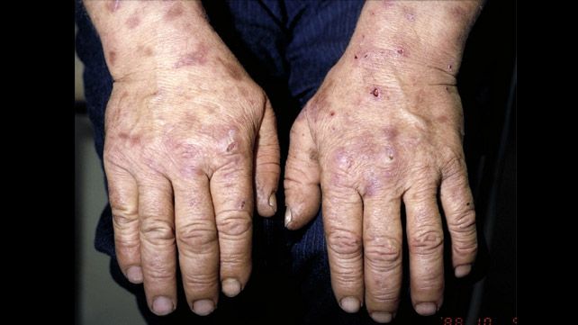
These porphyrias primarily manifest with skin lesions due to the accumulation of photosensitive porphyrins in the skin. When exposed to light, these porphyrins generate reactive oxygen species, leading to cellular damage.
- 1. Porphyria Cutanea Tarda (PCT):
- Etiology/Pathogenesis: The most common type of porphyria worldwide. Caused by a deficiency of uroporphyrinogen decarboxylase (UROD), the fifth enzyme in the heme pathway. Most cases are acquired (Type I), often triggered by factors like chronic alcohol use, hepatitis C infection, HIV, iron overload, estrogen therapy, and certain chemicals. A smaller proportion (Type II) is familial, inherited in an autosomal dominant manner, with symptomatic individuals still requiring environmental triggers [12]. The primary pathogenic mechanism is thought to involve inhibition of UROD by iron and specific inhibitors in the liver, leading to accumulation of uroporphyrin and heptacarboxyl porphyrin.
- Clinical Features: Characterized by chronic, blistering skin lesions on sun-exposed areas (hands, forearms, face). These vesicles and bullae heal slowly, often leading to milia, scarring, hyperpigmentation, and increased skin fragility. Hirsutism (especially on the face) is also common. Urine may be reddish-brown, particularly upon standing [12].
- Treatment: The primary goals are to reduce hepatic porphyrin production and eliminate associated risk factors. Therapeutic phlebotomy (venesection) to reduce iron stores is highly effective in acquired PCT. Low-dose chloroquine or hydroxychloroquine can also be used, which increases porphyrin excretion. Patients must avoid alcohol, iron supplements, and sunlight exposure. Treatment of underlying conditions like hepatitis C is also crucial [13].
- 2. Erythropoietic Protoporphyria (EPP) and X-linked Protoporphyria (XLP):
- Etiology/Pathogenesis:
- EPP: Autosomal recessive disorder, due to a partial deficiency of ferrochelatase (FECH), the final enzyme in the heme pathway. This leads to the accumulation of protoporphyrin in erythrocytes, plasma, and liver. Most patients have one inactivating FECH mutation and a common low-expression allele on the other chromosome [14].
- XLP: Caused by a gain-of-function mutation in the ALAS2 gene on the X chromosome, leading to increased ALAS2 activity and overproduction of protoporphyrin. This results in an X-linked dominant inheritance pattern [15].
- Clinical Features: Both EPP and XLP present with acute, painful photosensitivity shortly after sun exposure, typically in childhood. Unlike PCT, there are usually no blisters, but rather burning, stinging, itching, and redness that can mimic an allergic reaction. Chronic skin changes can include thickening, waxy appearance, and scarring around the nose and mouth. Liver complications (cholestasis, cholelithiasis, liver failure) can occur in a small percentage of patients due to insoluble protoporphyrin deposits [14, 15].
- Treatment: Strict sun protection is paramount. Oral beta-carotene can increase tolerance to light, though its efficacy is modest. Afamelanotide, a synthetic α-melanocyte stimulating hormone analog, is an approved therapy that increases melanin production and provides photoprotection. Acute pain is managed symptomatically. In cases of liver involvement, bile acid sequestrants may be used, and in severe cases, liver transplantation or red blood cell transfusions may be necessary [16].
- Etiology/Pathogenesis:
- 3. Congenital Erythropoietic Porphyria (CEP) / Günther’s Disease:
- Etiology/Pathogenesis: A very rare, severe, autosomal recessive disorder caused by a profound deficiency of uroporphyrinogen III synthase (UROS), the fourth enzyme in the heme pathway. This leads to the accumulation of highly photosensitive type I isomers of uroporphyrin and coproporphyrin in erythrocytes, plasma, and urine [17].
- Clinical Features: Symptoms typically begin in infancy, marked by severe photosensitivity leading to blistering, ulceration, and mutilating scars, especially on the face and extremities. Hirsutism, hemolytic anemia, splenomegaly, and reddish-brown urine (due to porphyrin excretion) are characteristic. Teeth may exhibit erythrodontia (reddish-brown discoloration) due to porphyrin deposition [17].
- Treatment: Treatment is challenging and aimed at reducing porphyrin levels and protecting from light. Strict sun protection, avoidance of trauma, and symptomatic management of skin lesions are essential. Chronic blood transfusions may suppress erythropoiesis and reduce porphyrin production. Bone marrow transplantation is the only curative treatment but carries significant risks. Experimental therapies are under investigation [18].
Conclusion
Porphyrias represent a fascinating and clinically significant group of disorders demanding a high index of suspicion for early diagnosis. Their diverse clinical manifestations, ranging from life-threatening neurovisceral attacks to debilitating chronic skin disease, underscore the critical role of heme synthesis in human physiology. Accurate diagnosis, often relying on biochemical testing of urine, plasma, and erythrocytes for porphyrins and their precursors, followed by genetic confirmation, is crucial for guiding specific therapeutic interventions. While some porphyrias can be effectively managed with lifestyle modifications and existing therapies, others remain challenging, highlighting the ongoing need for research into novel treatments to improve patient outcomes and quality of life.
References
[1] Balwani, M., & Desnick, R. J. (2012). The porphyrias: advances in diagnosis and treatment. Seminars in Liver Disease, 32(4), 316-324.
[2] Wang, B., & Bloomer, J. R. (2019). The porphyrias. In Sleisenger and Fordtran’s Gastrointestinal and Liver Disease (11th ed., pp. 1327-1336). Elsevier.
[3] Anderson, K. E., Sassa, S., Bishop, D. F., & Desnick, R. J. (2001). Disorders of Heme Biosynthesis: X-linked Sideroblastic Anemia and the Porphyrias. In C. R. Scriver, A. L. Beaudet, W. S. Sly, D. Valle, B. Childs, & B. Vogelstein (Eds.), The Metabolic and Molecular Bases of Inherited Disease (8th ed., pp. 2991-3068). McGraw-Hill.
[4] Simon, N. (2014). Porphyria: an overview. The Journal of Clinical Psychiatry, 75(5), e474-e478.
[5] Puy, H., Gouya, L., & Deybach, J. C. (2010). Porphyrias. The Lancet, 375(9718), 924-937.
[6] Hift, R. J., & Meissner, P. N. (2005). An approach to the diagnosis and management of the acute porphyrias. QJM: An International Journal of Medicine, 98(1), 1-12.
[7] Bissell, D. M., Wang, B., & Anderson, K. E. (2017). Porphyria. New England Journal of Medicine, 377(9), 862-872.
[8] Desnick, R. J., & Balwani, M. (2015). Acute Intermittent Porphyria. In R. A. Pagon et al. (Eds.), GeneReviews®. University of Washington, Seattle.
[9] Sardh, E., et al. (2019). Givosiran for acute hepatic porphyria. New England Journal of Medicine, 381(26), 2524-2536.
[10] Meissner, P. N., et al. (2013). Variegate Porphyria. In R. A. Pagon et al. (Eds.), GeneReviews®. University of Washington, Seattle.
[11] Schulze, T. G., & Badertscher, S. (2004). Hereditary Coproporphyria. In R. A. Pagon et al. (Eds.), GeneReviews®. University of Washington, Seattle.
[12] Gross, U., & Doss, M. O. (2000). Porphyria cutanea tarda: treatment principles based on molecular pathogenesis. Expert Opinion on Pharmacotherapy, 1(3), 441-452.
[13] Rocchi, E., et al. (2004). Porphyria cutanea tarda: a reappraisal. Journal of Hepatology, 41(4), 624-633.
[14] Whatley, S. D., et al. (2009). Erythropoietic protoporphyria and X-linked protoporphyria. Clinics in Dermatology, 27(5), 458-467.
[15] Toutain, P., et al. (2020). X-Linked Protoporphyria. In R. A. Pagon et al. (Eds.), GeneReviews®. University of Washington, Seattle.
[16] Langendonk, J. G., et al. (2015). Afamelanotide for erythropoietic protoporphyria. New England Journal of Medicine, 373(1), 48-59.
[17] Desnick, R. J., & Astrin, K. H. (2001). Congenital Erythropoietic Porphyria. In R. A. Pagon et al. (Eds.), GeneReviews®. University of Washington, Seattle.
[18] Singal, A. K., et al. (2019). Erythropoietic Porphyrias: Epidemiology, Pathogenesis, and Management. Dermatologic Clinics, 37(4), 543-561.


