Chromatography, derived from the Greek words “chroma” (color) and “graphein” (to write), is a powerful and versatile laboratory technique used for the separation, identification, and quantification of components in a mixture. First developed in 1903 by Mikhail Tsvet to separate plant pigments, it has evolved into an indispensable tool across diverse scientific disciplines, including chemistry, biochemistry, environmental science, and, critically, clinical diagnostics.
1. Principles of Chromatography
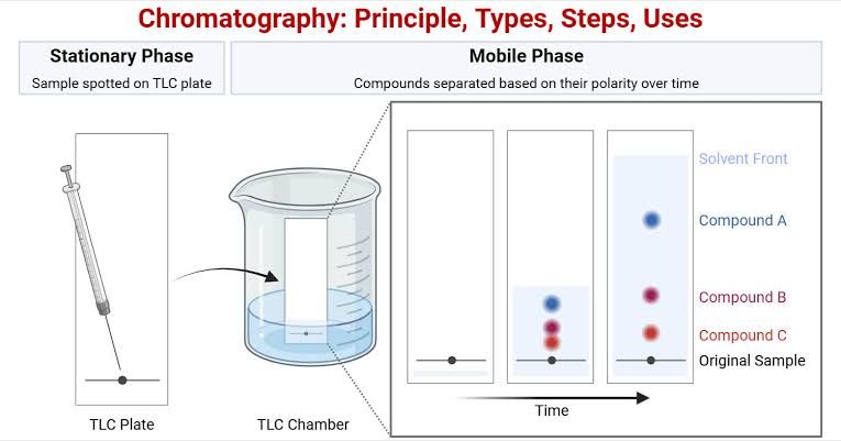
At its core, chromatography is a physical method of separation that relies on the differential distribution of components within a mixture between two immiscible phases: a stationary phase and a mobile phase.
1.1. The Stationary Phase: This phase is fixed in place and can be a solid, a liquid coated on a solid, or a gel. Its chemical properties (e.g., polarity, pore size, functional groups) dictate how strongly it interacts with the components of the mixture.
1.2. The Mobile Phase: This phase moves through or over the stationary phase, carrying the sample components with it. It can be a gas (Gas Chromatography, GC) or a liquid (Liquid Chromatography, LC), such as a solvent or a buffer.
1.3. Separation Mechanism: Differential Partitioning: When a sample mixture is introduced into the chromatographic system, its components begin to partition themselves between the stationary and mobile phases.
- Components that have a stronger affinity for the stationary phase (e.g., by adsorption, absorption, ion-exchange, or size exclusion) will spend more time interacting with it, thus moving more slowly through the system.
- Conversely, components with a stronger affinity for the mobile phase will be carried along more rapidly.
This differential interaction causes the components to separate from each other as they traverse the stationary phase. The time taken for a component to pass through the system is known as its retention time (tR) or, in planar chromatography, its retardation factor (Rf). These values, along with detector signals, allow for qualitative identification and quantitative analysis of the separated components.
2. Types of Chromatography
Chromatography encompasses a wide array of techniques, each optimized for specific applications based on the nature of the sample, the desired resolution, and the properties of the analytes. The primary classification is often based on the nature of the stationary and mobile phases, and the dominant separation mechanism.
2.1. Paper Chromatography (PC):
- Principle: A form of planar chromatography where the stationary phase is a sheet of filter paper (cellulose), and the mobile phase is a liquid solvent that moves through the paper by capillary action. Separation occurs based on differential partitioning between the water adsorbed onto the cellulose fibers and the organic solvent.
- Application: Primarily used for qualitative separation of amino acids, sugars, and pigments. It’s simple, inexpensive, and often used for educational purposes or preliminary screening.
2.2. Thin-Layer Chromatography (TLC):
- Principle: Similar to paper chromatography, but the stationary phase is a thin layer of adsorbent material (e.g., silica gel, alumina, cellulose) coated on an inert backing (glass, plastic, or aluminum foil). The mobile phase ascends the plate by capillary action.
- Application: Widely used for qualitative analysis, reaction monitoring, purity checks, drug screening, and initial compound identification in pharmaceutical, forensic, and clinical chemistry labs due to its speed, low cost, and good resolution.
2.3. Column Chromatography (General):
- Principle: The stationary phase is packed into a vertical glass or metal column, and the mobile phase is passed through it by gravity or pressure. This is a foundational technique from which more advanced column-based methods evolved.
- Application: Used for preparative separation and purification of larger quantities of compounds.
2.4. Gas Chromatography (GC):
- Principle: The mobile phase is an inert gas (e.g., helium, nitrogen, hydrogen), and the stationary phase is either a liquid coated on a solid support or a solid adsorbent, packed into a column or coated on the inner surface of a capillary. Components must be volatile or made volatile (derivatized) and thermally stable. Separation occurs based on boiling points and interactions with the stationary phase.
- Application: Ideal for volatile compounds like organic solvents, drugs of abuse, fatty acids, and gases. Often coupled with Mass Spectrometry (GC-MS) for definitive identification.
2.5. High-Performance Liquid Chromatography (HPLC):
- Principle: An advanced form of liquid chromatography where the mobile phase is pumped at high pressure through a column packed with very fine stationary phase particles. This high pressure forces the mobile phase through the column, resulting in high efficiency, speed, and resolution. HPLC can be further classified by the stationary phase polarity:
- Normal-Phase HPLC (NP-HPLC): Uses a polar stationary phase and a non-polar mobile phase.
- Reversed-Phase HPLC (RP-HPLC): Employs a non-polar stationary phase and a polar mobile phase (the most common type).
- Application: Extremely versatile, used for separating a wide range of non-volatile and thermally unstable compounds, including pharmaceuticals, proteins, peptides, vitamins, metabolites, and environmental contaminants. Often coupled with Mass Spectrometry (HPLC-MS) or UV-Vis detection.
2.6. Ion-Exchange Chromatography (IEC):
- Principle: Separation is based on the reversible electrostatic interaction between charged molecules (ions) in the sample and oppositely charged functional groups covalently bound to the stationary phase resin. The mobile phase is a buffer whose pH or ionic strength can be altered to elute the bound molecules.
- Application: Primarily used for separating charged molecules such as proteins, peptides, amino acids, nucleotides, and inorganic ions. Clinical applications include hemoglobin variant analysis and purification of therapeutic proteins.
2.7. Size-Exclusion Chromatography (SEC) / Gel Filtration Chromatography:
- Principle: Separation occurs based on the molecular size or hydrodynamic volume of components. The stationary phase consists of porous beads with pores of a defined size range. Larger molecules are excluded from the pores and elute first, while smaller molecules enter the pores, taking a longer, more tortuous path and eluting later.
- Application: Used for separating macromolecules like proteins, nucleic acids, and polysaccharides, determining molecular weight, and desalinating or buffer exchanging samples.
2.8. Affinity Chromatography (AC):
- Principle: The most specific chromatographic technique. Separation relies on a reversible and specific biological interaction between a target molecule in the sample and a highly specific ligand (e.g., antibody, enzyme substrate, receptor) covalently immobilized on the stationary phase. Unbound molecules wash through, while the target molecule binds, and is then eluted by altering conditions (e.g., pH, salt concentration).
- Application: Highly effective for purifying specific biomolecules from complex mixtures, such as antibodies, recombinant proteins, and receptors.
3. Interpretation of Clinical Conditions and Investigations Related to Use in Chromatography
Chromatography plays a pivotal role in clinical diagnostics, disease monitoring, and therapeutic interventions. Its ability to separate and quantify specific biomolecules, drugs, and metabolites in biological samples (blood, urine, CSF) provides crucial information for patient care.
3.1. Metabolic Disorders:
- Application: GC-MS and LC-MS/MS are routinely used to screen for inborn errors of metabolism (IEMs).
- Interpretation:
- Aminoacidopathies (e.g., Phenylketonuria – PKU): LC-MS/MS can quantify specific amino acids (e.g., elevated phenylalanine in PKU) in dried blood spots from newborns. An abnormal chromatogram showing elevated levels of a specific amino acid, compared to established reference ranges, indicates a potential metabolic block.
- Organic Acidemias (e.g., Methylmalonic Acidemia): GC-MS analysis of urine provides a “metabolic fingerprint” of organic acids. The presence of abnormal peaks or significantly elevated levels of specific organic acids (e.g., methylmalonic acid, propionic acid) points to enzymatic deficiencies that impair metabolic pathways.
- Clinical Relevance: Early detection allows for timely dietary intervention or treatment, preventing severe developmental delays and neurological damage.
3.2. Therapeutic Drug Monitoring (TDM):
- Application: HPLC and LC-MS/MS are commonly used to measure circulating levels of therapeutic drugs.
- Interpretation:
- Monitoring drugs with narrow therapeutic windows (e.g., anticonvulsants like phenytoin, immunosuppressants like cyclosporine, antibiotics like vancomycin).
- Chromatograms are analyzed for the drug’s peak area or height, which is then quantified against a calibration curve. The measured concentration is compared to the established therapeutic range for that drug.
- Clinical Relevance: Ensures optimal drug efficacy while minimizing toxicity. Sub-therapeutic levels may indicate non-compliance or malabsorption, while supra-therapeutic levels suggest potential toxicity or impaired clearance.
3.3. Drug Screening and Toxicology:
- Application: GC-MS and LC-MS/MS are the gold standards for detecting illicit drugs, prescription drug abuse, and poisons in biological fluids (urine, blood, hair).
- Interpretation:
- Chromatograms are meticulously examined for the presence of specific drug peaks or their metabolites. Retention times are used for presumptive identification, while mass spectral data provides definitive confirmation and structural elucidation.
- Quantification of drug levels can help determine the extent of exposure or overdose.
- Clinical Relevance: Essential for forensic investigations, emergency toxicology, monitoring drug rehabilitation programs, and managing suspected poisoning cases.
3.4. Hemoglobinopathies:
- Application: HPLC is widely used for the separation and quantification of normal and abnormal hemoglobin variants.
- Interpretation:
- HbA1c Monitoring for Diabetes: HPLC accurately quantifies glycated hemoglobin (HbA1c), which reflects average blood glucose levels over the preceding 2-3 months. Elevated HbA1c percentages indicate poor glycemic control.
- Sickle Cell Disease and Thalassemia Screening: HPLC chromatograms can identify and quantify various hemoglobin variants (e.g., HbS, HbC, HbE, HbF). An abnormal pattern (e.g., predominant HbS peak with reduced HbA) is indicative of specific hemoglobinopathies.
- Clinical Relevance: Crucial for managing diabetes, diagnosing inherited blood disorders, and carrier screening.
3.5. Nutritional Status Assessment:
- Application: HPLC is employed to quantify levels of various vitamins (e.g., fat-soluble vitamins A, D, E, K; water-soluble vitamins B and C) and their metabolites in plasma or serum.
- Interpretation:
- Chromatograms show distinct peaks for each vitamin. Peak areas are quantified and compared to established normal ranges.
- Low levels may indicate dietary deficiency, malabsorption, or increased requirements. Elevated levels can point to toxicity (especially for fat-soluble vitamins).
- Clinical Relevance: Helps diagnose and monitor vitamin deficiencies or toxicities, particularly in patients with malabsorption syndromes, chronic diseases, or those on specialized diets.
In conclusion, chromatography is a multifaceted and indispensable analytical technique. Its foundational principles of differential partitioning, combined with the diverse array of specialized methods, enable the precise separation and identification of countless compounds. In clinical settings, the interpretation of chromatographic data provides invaluable insights into metabolic health, drug efficacy and toxicity, and the presence of inherited conditions, thereby guiding diagnosis, treatment, and ongoing patient management. The continuous evolution of chromatographic technologies promises even greater precision and broader applications in future clinical investigations.


